
Ganesh Kumar S, Kalimuthu K, Solomon Robinson David Jebakumar, Vimalan J
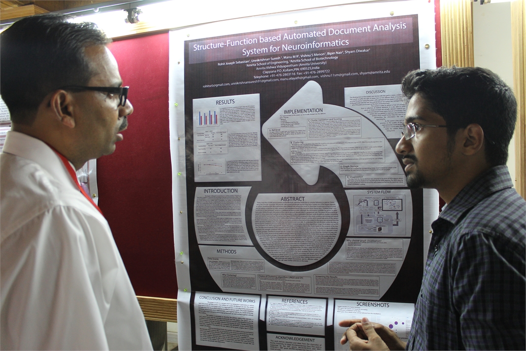
Molecular and physicochemical characterization of rhamnolipid biosurfactant produced by Pseudomonas sp JSK6
Interest in microbial surfactants like rhamnolipids has been progressively increasing in recent years due to their diversity, biodegradability and possibility of large scale production (Pinzon et al. 2013). However, traditional engineering by random and targeted genetic alteration, process design and recombinant strategies in rhamnolipid production were not studied in detail. Based on the environmental conditions, wide diversity of rhamnolipid congeners and homologues was produced in various bacterial strains (Abdel-Mawgoud et al. 2010). In this study, a biosurfactant producing bacterial strain, Pseudomonas sp. JSK6 was isolated from hydrocarbons contaminated sites at Madurai district of South India. The isolate produced mixture of both mono and di-rhamnolipids with excellent surfactant properties. The critical micelle concentration (CMC) of the produced rhamnolipids was 30 mg/L. The emulsification index of 56.4% with diesel and 55.3% with kerosene was quite stable after 24 h. The culture condition optimisation showed that highest rhamnolipid (1.6 g/L) production was achieved at pH 7 and 35◦C. The rhamnolipid was stable over a wide range of temperature (upto 100◦C) and pH (upto 10). The enzyme rhamnosyltransferase-1 which is responsible for biosynthesis of mono-rhamnolipid is encoded by the rhIAB genes, which are organized in rhlABRI operon. From the isolate JSK6, mono-rhamnolipid encoding gene cluster rhlABRI (4.3 kb) was successfully amplified and sequenced by primer walking. The sequence analysis showed that the rhlABRI gene cluster was perfectly organized in the strain JSK6. The rhamnolipid produced by strain JSK6 was analysed in NMR and LC-ESI-MS for structural characterisation. The chemical shifts in 1HNMR (0.86 ppm, 1.267 ppm, 2.546 ppm, 4.134ppm and 5.427 ppm) and 13CNMR (14.466ppm, 23.004–34.951ppm, 171.833ppm and 174.056ppm) and ESI-MS analysis suggested that extracted biosurfactant was present as mono-rhamnolipid and di-rhamnolipid as ten different homologues. This study concluded that the isolate JSK6 is a potential strain with capabilities to produce rhamnolipid biosurfactants with unique physicochemical properties. Cloning of rhlABRI gene cluster of strain JSK6 in a suitable expression system for large scale production is under progress.
 Kartik Iyer
Assistant Professor, School of Biotechnology, Amrita University
 Shyam Diwakar, Ph.D.
Assistant Professor, Amrita School of Biotechnology

Dr. Bipin Nair,
Dean-Biotechnology, Amrita University
 Suryaprakash Sambhara, DVM, Ph.D
Chief, Immunology Section, Influenza Division, CDC, Atlanta, USA
Making sense of pathogen sensors of Innate Immunity: Utility of their ligands as antiviral agents and adjuvants for vaccines.
Currently used antiviral agents act by inhibiting viral entry, replication, or release of viral progeny. However, recent emergence of drug-resistant viruses has become a major public health concern as it is limiting our ability to prevent and treat viral diseases. Furthermore, very few antiviral agents with novel modes of action are currently in development. It is well established that the innate immune system is the first line of defense against invading pathogens. The recognition of diverse pathogen-associated molecular patterns (PAMPs) is accomplished by several classes of pattern recognition receptors (PRRs) and the ligand/receptor interactions trigger an effective innate antiviral response. In the past several years, remarkable progress has been made towards understanding both the structural and functional nature of PAMPs and PRRs. As a result of their indispensable role in virus infection, these ligands have become potential pharmacological agents against viral infections. Since their pathways of action are evolutionarily conserved, the likelihood of viruses developing resistance to PRR activation is diminished. I will discuss the recent developments investigating the potential utility of the ligands of innate immune receptors as antiviral agents and molecular adjuvants for vaccines.


 Jaap Heringa, Ph.D.
Director & Professor of Bioinformatics, IBIVU VU University Amsterdam, The Netherlands
Modeling strategy based on Petri-nets
In my talk I will introduce a formal modeling strategy based on Petri-nets, which are a convenient means of modeling biological processes. I will illustrate the capabilities of Petri-nets as reasoning vehicles using two examples: Haematopoietic stem cell differentiation in mice, and vulval development in C. elegance. The first system was modeled using a Boolean implementation, and the second using a coarse-grained multi-cellular Petri-net model. Concepts such as the model state space, attractor states, and reasoning to adapt the model to the biological reality will be discussed.
 Shantikumar Nair, Ph.D.
Professor & Director, Amrita Center for Nanosciences & Molecular Medicine, Amrita University, India
Spatially Distributed and Hierarchical Nanomaterials in Biotechnology
Although nano materials are well investigated in biotechnology in their zero-, one- and two-dimensional forms, three-dimensional nanomaterials are relatively less investigated for their biological applications. Three dimensional nano materials are much more complex with several structural and hierarchical variables controlling their mechanical, chemical and biological functionality. In this talk examples are given of some complex three dimensional systems including, scaffolds, aggregates, fabrics and membranes. Essentially three types of hierarchies are considered: one-dimensional hierarchy, two-dimensional hierarchy and three-dimensional hierarchy each giving rise to unique behaviors.

 Nader Pourmand, Ph.D.
Director, UCSC Genome Technology Center,University of California, Santa Cruz
Biosensor and Single Cell Manipulation using Nanopipettes
Approaching sub-cellular biological problems from an engineering perspective begs for the incorporation of electronic readouts. With their high sensitivity and low invasiveness, nanotechnology-based tools hold great promise for biochemical sensing and single-cell manipulation. During my talk I will discuss the incorporation of electrical measurements into nanopipette technology and present results showing the rapid and reversible response of these subcellular sensors to different analytes such as antigens, ions and carbohydrates. In addition, I will present the development of a single-cell manipulation platform that uses a nanopipette in a scanning ion-conductive microscopy technique. We use this newly developed technology to position the nanopipette with nanoscale precision, and to inject and/or aspirate a minute amount of material to and from individual cells or organelle without comprising cell viability. Furthermore, if time permits, I will show our strategy for a new, single-cell DNA/ RNA sequencing technology that will potentially use nanopipette technology to analyze the minute amount of aspirated cellular material.
 Rajgopal Srinivasan, Ph.D.
Principal Scientist & Head Bio IT R&D, TCS Innovation Labs, India
Interpretation of Genomic Variation – Identifying Rare Variations Leading to Disease
Genome sequencing technologies are generating an abundance of data on human genetic variations. A big challenge lies in interpreting the functional relevance of such variations, especially in clinical settings. A first step in understanding the clinical relevance of genetic variations is to annotate the variants for region of occurrence, degree of conservation both within and across species, pattern of variation across related individuals, novelty of the variation and know effects of related variations. Several tools already exist for this purpose. However, these tools have their strengths and weaknesses. A second issue is the development of algorithms, which, given a rich annotation of variants are able to prioritize the variants as being relevant to the phenotype under investigation.
In my talk I will detail work that has been done in our labs to address both of the above problems. I will also illustrate the application of these tools that helped identify a rare mutation in the ATM gene leading to a diagnosis of AT in two infants.
 D. Narasimha Rao, Ph.D.
Professor, Dept of Biochemistry, Indian Institute of Science, Bangalore, India
Genomics of Restriction-Modification Systems
Restriction endonucleases occur ubiquitously among procaryotic organisms. Up to 1% of the genome of procaryotic organisms is taken up by the genes for these enzymes. Their principal biological function is the protection of the host genome against foreign DNA, in particular bacteriophage DNA. Restriction-modification (R-M) systems are composed of pairs of opposing enzyme activities: an endonuclease and a DNA methyltransferase (MTase). The endonucleases recognise specific sequences and catalyse cleavage of double-stranded DNA. The modification MTases catalyse the addition of a methyl group to one nucleotide in each strand of the recognition sequence using S-adenosyl-L-methionine (AdoMet) as the methyl group donor. Based on their molecular structure, sequence recognition, cleavage position and cofactor requirements, R-M systems are generally classified into three groups. In general R-M systems restrict unmodified DNA, but there are other systems that specifically recognise and cut modified DNA. More than 3500 restriction enzymes have been discovered so far. With the identification and sequencing of a number of R-M systems from bacterial genomes, an increasing number of these have been found that do not seem to fit into the conventional classification.
It is well documented that restriction enzyme genes always lie close to their cognate methyltransferase genes. Analysis of the bacterial and archaeal genome sequences shows that MTase genes are more common than one would have expected on the basis of previous biochemical screening. Frequently, they clearly form part of a R-M system, because the adjacent open reading frames (ORFs) show similarity to known restriction enzyme genes. Very often, though, the adjacent ORFs have no homologs in the GenBank and become candidates either for restriction enzymes with novel specificities or for new examples of previously uncloned specificities. Sequence-dependent modification and restriction forms the foundation of defense against foreign DNAs and thus RM systems may serve as a tool of defense for bacterial cells. RM systems however, sometimes behave as discrete units of life, and any threat to their maintenance, such as a challenge by a competing genetic element can lead to cell death through restriction breakage in the genome, thus providing these systems with a competitive advantage. The RM systems can behave as mobile-genetic elements and have undergone extensive horizontal transfer between genomes causing genome rearrangements. The capacity of RM systems to act as selfish, mobile genetic elements may underlie the structure and function of RM enzymes.
The similarities and differences in the different mechanisms used by restriction enzymes will be discussed. Although it is not clear whether the majority of R-M systems are required for the maintenance of the integrity of the genome or whether they are spreading as selfish genetic elements, they are key players in the “genomic metabolism” of procaryotic organisms. As such they deserve the attention of biologists in general. Finally, restriction enzymes are the work horses of molecular biology. Understanding their enzymology will be advantageous to those who use these enzymes, and essential for those who are devoted to the ambitious goal of changing the properties of these enzymes, and thereby make them even more useful.

 Ajay Shah, Ph.D.
 Michelle Hermiston, MD, Ph.D.
Assistant Professor, Department of Pediatrics University of California San Francisco, USA
Interrogating Signaling Networks at the Single Cell Level In Primary Human Patient Samples
Multiparameter phosphoflow cytometry is a highly sensitive proteomic approach that enables monitoring of biochemical perturbations at the single cell level. By combining antisera to cell surface markers and key intracellular proteins, perturbations in signaling networks, cell survival and apoptosis mediators, cell cycle regulators, and/or modulators of other cellular processes can be analyzed in a highly reproducible and sensitive manner in the basal state and in response to stimulation or drug treatment. Advantages of this approach include the ability to identify the biochemical consequences of genetic and/or epigenetic changes in small numbers of cells, to map potential interplay between various signaling networks simultaneously in a single cell, and to interrogate potential mechanisms of drug resistance or response in a primary patient sample. Application of this technology to patients with acute lymphoblastic leukemia or the autoimmune disease systemic lupus erythematosus (SLE) will be discussed.

Jaydeep Unni, Ph.D.
Sr. Project Manager, Robert Bosch Healthcare Systems, Palo Alto, CA
Remote Patient Monitoring – Challenges and Opportunities
Remote Patient Monitoring (RPM) is gaining importance and acceptance with rising number of chronic disease conditions and with increase in the aging population. As instances of Heart diseases, Diabetes etc are increasing the demand for these technologies are increasing. RPM devices typically collect patient vital sign data and in some case also patient responses to health related questions. Thus collected data is then transmitted through various modalities (wireless/Bluetooth/cellular) to Hospitals/Doctor’s office for clinical evaluation. With these solutions Doctors are able to access patient’s vital data ‘any time any where’ thus enabling them to intervene on a timely and effective manner. For older adult population chronic disease management, post-acute care management and safety monitoring are areas were RPM finds application. That said, there are significant challenges in adoption of Remote Patient Monitoring including patient willingness and compliance for adoption, affordability, availability of simpler/smarter technology to mention a few. But experts contend that if implemented correctly Remote Patient Monitoring can contain healthcare expenditure by reducing avoidable hospitalization while greatly improving quality of care.
 Kal Ramnarayan, Ph.D.
Co-founder President & Chief Scientific Officer, Sapient Discovery, San Diego, CA, USA
A cost-effective approach to Protein Structure-guided Drug Discovery: Aided by Bioinformatics, Chemoinformatics and computational chemistry
With the mapping of the human genome completed almost a decade ago, efforts are still underway to understand the gene products (i.e., proteins) in the human biological and disease pathways. Deciphering such information is very important for the discovery and development of small molecule drugs as well as protein therapeutics for various human diseases for which no cure exists. As an example, with more than 500 members, the kinase family of protein targets continues to be an important and attractive class for drug discovery. While how many of the members in this family are actually druggable is still to be established, there are several ongoing efforts on this class of proteins across a broad spectrum of disease categories. Even though in general the protein structural topology might looks similar, there are issues with respect selectivity of identified small molecule inhibitors when, the lead molecule discovery is carried out at the ATP binding site. As an added complexity, allosteric modulators are needed for some of the members, but the actual site for such modulation on the protein target can not resolved with uncertainty. In this presentation we will describe a bioinformatics and computational based platform for small molecule discovery for protein targets that are involved in protein-protein interactions as well as targets like kinases and phosphatases. We will describe a computational approach in which we have used an informatics based platform with several hundred kinases to sort through in silico and identify inhibitors that are likely to be highly selective in the lead generation phase. We will discuss the implication of this approach on the drug discovery of the kinase and phosphatase classes in general and independent of the disease category.
 Shigeki Miyamoto, Ph.D.
Professor, McArdle Laboratory for Cancer Research – UW Carbone Cancer Center
Department of Oncology, School of Medicine and Public Health
University of Wisconsin-Madison
“Inside-out” NF-κB signaling in cancer and other pathologies
The NF-κB/Rel family of transcription factors contributes to critical cellular processes, including immune, inflammatory and cell survival responses. As such, NF-κB is implicated in immunity-related diseases, as well as multiple types of human malignancies. Indeed, genetic alterations in the NF-κB signaling pathway are frequently observed in multiple human malignancies. NF-κB is normally kept inactive in the cytoplasm by inhibitor proteins. Extracellular ligands can induce the release of NF-κB from the inhibitors to allow its migration into the nucleus to regulate a variety of target genes. NF-κB activation is also induced in response to multiple stress conditions, including those induced by DNA-damaging anticancer agents. Although precise mechanisms are still unclear, research from our group has revealed a unique nuclear-to-cytoplasmic signaling pathway. In collaboration with bioengineers, clinicians and pharmaceutical industry, our lab has developed new methods to analyze primary cancer patient samples and identified several compounds with different mechanisms that mitigate this cell survival pathway. Further contributions from other labs have also revealed additional mechanisms and molecular players in this “inside-out” signaling pathway and expanded its role in other physiological and pathological processes, including B cell development, premature aging and therapy resistance of certain cancers. Our own new findings, along with these recent developments in the field, will be highlighted.

 Prashanth Athri, Ph.D.
Senior Specialist, Strand Life Sciences, Bengaluru, India
Rare disease diagnostic platform
At Strand, genomic sequencing combined with bioinformatic analysis have provided discriminative diagnosis in the case of rare genetic disorders. Inspired by these cases, we are building an integrated software that combines curated literature content and bioinformatics databases with a clinically oriented user interface to substantially compress time taken to determine likely candidate genetic variants in a Diagnostic Odyssey. At the back end we employ various algorithms that systematically query our diverse knowledgebase to provide the clinicians a comprehensive, and possibly multidimensional, annotation of the variant in the context of disease.
 Sneh Anand, Ph.D.
Professor, Center for Biomedical Engineering, IIT-Delhi, India
Interdisciplinary Research Outcome of Biomedical Engineering
Natural science is an engineering marvel. All innovations in health care technology have been inspired by biological systems. A joint venture of the two premier Institutes has facilitated research in Biomedical engineering. Over the years the Centre is a premier in the country with global recognition. This interdisciplinary base platform has lead to several innovative technologies which have been patented and validated by clinicians as well. The R&D contributions in mass health care, diagnostics, therapeutics and rehabilitation reinforcement. Graduate exposure to the field can enhance creativity among graduates from all engineering disciplines.
 V. Nagaraja Ph.D.
Professor, Indian Institute of Science, Bengaluru, India
Perturbation of DNA topology in mycobacteria
To maintain the topological homeostasis of the genome in the cell, DNA topoisomerases catalyse DNA cleavage, strand passage and rejoining of the ends. Thus, although they are essential house- keeping enzymes, they are the most vulnerable targets; arrest of the reaction after the first trans-esterification step leads to breaks in DNA and cell death. Some of the successful antibacterial or anticancer drugs target the step ie arrest the reaction or stabilize the topo -DNA covalent complex. I will describe our efforts in this direction – to target DNA gyrase and also topoisomerase1 from mycobacteria. The latter, although essential, has no inhibitors described so far. The new inhibitors being characterized are also used to probe topoisomerase control of gene expression.
In the biological warfare between the organisms, a diverse set of molecules encoded by invading genomes target the above mentioned most vulnerable step of topoisomerase reaction, leading to the accumulation of double strand breaks. Bacteria, on their part appear to have developed defense strategies to protect the cells from genomic double strand breaks. I will describe a mechanism involving three distinct gyrase interacting proteins which inhibit the enzyme in vitro. However, in vivo all these topology modulators protect DNA gyrase from poisoning effect by sequestering the enzyme away from DNA.
Next, we have targeted a topology modulator protein, a nucleoid associated protein(NAP) from Mycobacterium tuberculosis to develop small molecule inhibitors by structure based design. Over expression of HU leads to alteration in the nucleoid architecture. The crystal structure of the N-terminal half of HU reveals a cleft that accommodates duplex DNA. Based on the structural feature, we have designed inhibitors which bind to the protein and affect its interaction with DNA, de-compact the nucleoid and inhibit cell growth. Chemical probing with the inhibitors reveal the importance of HU regulon in M.tuberculosis.
 R. Manjunath, Ph.D.
Associate Professor, Dept of Biochemistry, Indian Institute of Science, Bengaluru, India
REGULATION OF THE MHC COMPLEX AND HLA SOLUBILISATION BY THE FLAVIVIRUS, JAPANESE ENCEPHALITIS VIRUS
Viral encephalitis caused by Japanese encephalitis virus (JEV) and West Nile Virus (WNV) is a mosquito-borne disease that is prevalent in different parts of India and other parts of South East Asia. JEV is a positive single stranded RNA virus that belongs to the Flavivirus genus of the family Flaviviridae. The genome of JEV is about 11 kb long and codes for a polyprotein which is cleaved by both host and viral encoded proteases to form 3 structural and 7 non-structural proteins. It is a neurotropic virus which infects the central nervous system (CNS) and causes death predominantly in newborn children and young adults. JEV follows a zoonotic life-cycle involving mosquitoes and vertebrate, chiefly pigs and ardeid birds, as amplifying hosts. Humans are infected when bitten by an infected mosquito and are dead end hosts. Its structural, pathological, immunological and epidemiological aspects have been well studied. After entry into the host following a mosquito bite, JEV infection leads to acute peripheral neutrophil leucocytosis in the brain and leads to elevated levels of type I interferon, macrophage-derived chemotactic factor, RANTES,TNF-α and IL-8 in the serum and cerebrospinal fluid.
Major Histocompatibility Complex (MHC) molecules play a very important role in adaptive immune responses. Along with various classical MHC class I molecules, other non-classical MHC class I molecules play an important role in modulating innate immune responses. Our lab has shown the activation of cytotoxic T-cells (CTLs) during JEV infection and CTLs recognize non-self peptides presented on MHC molecules and provide protection by eliminating infected cells. However, along with proinflammatory cytokines such as TNFα, they may also cause immunopathology within the JEV infected brain. Both JEV and WNV, another related flavivirus have been shown to increase MHC class I expression. Infection of human foreskin fibroblast cells (HFF) by WNV results in upregulation of HLA expression. Data from our lab has also shown that JEV infection upregulates classical as well as nonclassical (class Ib) MHC antigen expression on the surface of primary mouse brain astrocytes and mouse embryonic fibroblasts.
There are no reports that have discussed the expression of these molecules on other cells like endothelial and astrocyte that play an important role in viral invasion in humans. We have studied the expression of human classical class I molecules HLA-A, -B, -C and the non-classical HLA molecules, HLA-E as well as HLA-F in immortalized human brain microvascular endothelial cells (HBMEC), human endothelial cell line (ECV304), human glioblastoma cell line (U87MG) and human foreskin fibroblast cells (HFF). Nonclassical MHC molecules such as mouse Qa-1b and its human homologue, HLA-E have been shown to be the ligand for the inhibitory NK receptor, NKG2A/CD94 and may bridge innate and adaptive immune responses. We show that JEV infection of HBMEC and ECV 304 cells upregulates the expression of HLA-A, and –B antigens as well as HLA-E and HLA-F. Increased expression of total HLA-E upon JEV infection was also observed in other human cell lines as well like, human amniotic epithelial cells, AV-3, FL and WISH cells. Further, we show for the first time that soluble HLA-E (sHLA-E) was released from infected ECV and HBMECs. In contrast, HFF cells showed only upregulation of cell-surface HLA-E expression while U87MG, a human glioblastoma cell line neither showed any cell-surface induction nor its solubilization. This shedding of sHLA-E was found to be dependent on matrix metalloproteinase (MMP) and an important MMP, MMP-9 was upregulated during JEV infection. Treatment with IFNγ resulted in the shedding of sHLA-E from ECV as well as U87MG but not from HFF cells. Also, sHLA-E was shed upon treatment with IFNβ and both IFNβ and TNFα, when present together caused an additive increase in the shedding of sHLA-E. HLA-E is an inhibitory ligand for CD94/NKG2A receptor of Natural Killer cells. Thus, MMP mediated solubilization of HLA-E from infected endothelial cells may have important implications in JEV pathogenesis including its ability to compromise the blood brain barrier.


 Karmeshu, Ph.D.
Dean & Professor, School of Computer & Systems Sciences & School of Computational & Integrative Sciences, Jawaharlal Nehru University, India.
Interspike Interval Distribution of Neuronal Model with distributed delay: Emergence of unimodal, bimodal and Power law
The study of interspike interval distribution of spiking neurons is a key issue in the field of computational neuroscience. A wide range of spiking patterns display unimodal, bimodal ISI patterns including power law behavior. A challenging problem is to understand the biophysical mechanism which can generate the empirically observed patterns. A neuronal model with distributed delay (NMDD) is proposed and is formulated as an integro-stochastic differential equation which corresponds to a non-markovian process. The widely studied IF and LIF models become special cases of this model. The NMDD brings out some interesting features when excitatory rates are close to inhibitory rates rendering the drift close to zero. It is interesting that NMDD model with gamma type memory kernel can also account for bimodal ISI pattern. The mean delay of the memory kernels plays a significant role in bringing out the transition from unimodal to bimodal ISI distribution. It is interesting to note that when a collection of neurons group together and fire together, the ISI distribution exhibits power law.
 Sharmila Mande, Ph.D.
Principal Scientist and Head, Bio Sciences R&D, TCS Innovation Labs, Pune
Gut microbiome and health: Moving towards the new era of translational medicine
The microbes inhabiting our body outnumber our own cells by a factor of 10. The genomes of these microbes, called the ‘second genome’ are therefore expected to have great influence on our health and well being. The emerging field of metagenomics is rapidly becoming the method of choice for studying the microbial community (called microbiomes) present in various parts of the human body. Recent studies have implicated the role of gut microbiomes in several diseases and disorders. Studies have indicated gut microbiome’s role in nutrient absorption, immuno-modulation motor-response, and other key physiological processes. However, our understanding of the role of gut microbiota in malnutrition is currently incomplete. Exploration of these aspects are likely to help in understanding the microbial basis for several physiological disorders associated with malnutrition (eg, increased susceptibility to diarrhoeal pathogens) and may finally aid in devising appropriate probiotic strategies addressing this menace. A metagenomic approach was employed for analysing the differences between gut microbial communities obtained from malnourished and healthy children. Results of the analysis using TCS’ ‘Metagenomic Analysis Platform’ were discussed in detail during my talk.
 Seeram Ramakrishna, Ph.D.
Director, Center for Nanofibers & Nanotechnology, National University of Singapore
Biomaterials: Future Perspectives
From the perspective of thousands of years of history, the role of biomaterials in healthcare and wellbeing of humans is at best accidental. However, since 1970s with the introduction of national regulatory frameworks for medical devices, the biomaterials field evolved and reinforced with strong science and engineering understandings. The biomaterials field also flourished on the backdrop of growing need for better medical devices and medical treatments, and sustained investments in research and development. It is estimated that the world market size for medical devices is ~300 billion dollars and for biomaterials it is ~30 billion dollars. Healthcare is now one of the fastest growing sectors worldwide. Legions of scientists, engineers, and clinicians worldwide are attempting to design and develop newer medical treatments involving tissue engineering, regenerative medicine, nanotech enabled drug delivery, and stem cells. They are also engineering ex-vivo tissues and disease models to evaluate therapeutic drugs, biomolecules, and medical treatments. Engineered nanoparticles and nanofiber scaffolds have emerged as important class of biomaterials as many see them as necessary in creating suitable biomimetic micro-environment for engineering and regeneration of various tissues, expansion & differentiation of stem cells, site specific controlled delivery of biomolecules & drugs, and faster & accurate diagnostics. This lecture will capture the progress made thus far in pre-clinical and clinical studies. Further this lecture will discuss the way forward for translation of bench side research into the bed side practice. This lecture also seeks to identify newer opportunities for biomaterials beyond the medical devices.

 Manzoor K, Ph.D.
Professor, Centre for Nanoscience & Molecular Medicine, Amrita University
Targeting aberrant cancer kinome using rationally designed nano-polypharmaceutics
Manzoor Koyakutty, Archana Ratnakumary, Parwathy Chandran, Anusha Ashokan, and Shanti Nair
`War on Cancer’ was declared nearly 40 years ago. Since then, we made significant progress on fundamental understanding of cancer and developed novel therapeutics to deal with the most complex disease human race ever faced with. However, even today, cancer remains to be the unconquered `emperor of all maladies’. It is well accepted that meaningful progress in the fight against cancer is possible only with in-depth understanding on the molecular mechanisms that drives its swift and dynamic progression. During the last decade, emerging new technologies such as nanomedicine could offer refreshing life to the `war on cancer’ by way of providing novel methods for molecular diagnosis and therapy.
In the present talk, we discuss our approaches to target critically aberrant cancer kinases using rationally designed polymer-protein and protein-protein core-shell nanomedicines. We have used both genomic and proteomic approaches to identify many intimately cross-linked and complex aberrant protein kinases behind the drug resistance and uncontrolled proliferation of refractory leukemic cells derived from patients. Small molecule inhibitors targeted against oncogenic pathways in these cells were found ineffective due to the involvement of alternative survival pathways. This demands simultaneous inhibition more than one oncogenic kinases using poly-pharmaceutics approach. For this, we have rationally designed core-shell nanomedicines that can deliver several small molecules together for targeting multiple cancer signalling. We have also used combination of small molecules and siRNA for combined gene silencing together with protein kinase inhibition in refractory cancer cells. Optimized nanomedicines were successfully tested in patient samples and found enhanced cytotoxicity and molecular specificity in drug resistant cases.
Nano-polypharmaceutics represents a new generation of nanomedicines that can tackle multiple cancer mechanisms simultaneously. Considering the complexity of the disease, such therapeutic approaches are not simply an advantage, but indispensable.
Acknowledgements:
We thank Dept. of Biotechnology and Dept. Of Science and Technology,Govt. of India for the financial support through `Thematic unit of Excellence in Medical NanoBiotechnology’ and `Nanomedicine- RNAi programs’.

 Lalitha Subramanian, Ph.D.
Chief Scientific Officer & VP, Services at Scienomics, USA
Nanoscale Simulations – Tackling Form and Formulation Challenges in Drug Development and Drug Delivery
Lalitha Subramanian, Dora Spyriouni, Andreas Bick, Sabine Schweizer, and Xenophon Krokidis Scienomics
The discovery of a compound which is potent in activity against a target is a major milestone in Pharmaceutical and Biotech industry. However, a potent compound is only effective as a therapeutic agent when it can be administered such that the optimal quantity is transported to the site of action at an optimal rate. The active pharmaceutical ingredient (API) has to be tested for its physicochemical properties before the appropriate dosage form and formulation can be designed. Some of the commonly evaluated parameters are crystal forms and polymorphs, solubility, dissolution behavior, stability, partition coefficient, water sorption behavior, surface properties, particle size and shape, etc. Pharmaceutical development teams face the challenge of quickly and efficiently determining a number of properties with small quantities of the expensive candidate compounds. Recently the trend has been to screen these properties as early as possible and often the candidate compounds are not available in sufficient quantities. Increasingly, these teams are leveraging nanoscale simulations similar to those employed by drug discovery teams for several decades. Nanoscale simulations are used to predict the behavior using very little experimental data and only if this is promising further experiments are done. Another aspect where nanoscale simulations are being used in drug development and drug delivery is to get insights into the behavior of the system so that process failures can be remediated and formulation performance can be improved. Thus, the predictive screening and the in-depth understanding leads to experimental efficiency resulting in far-reaching business impacts.
With specific examples, this talk will focus on the different types of nanoscale simulations used to predict properties of the API in excipients and also provide insight into system behavior as a function of shelf life, temperature, mechanical stress, etc.
 Satheesh Babu T. G., Ph.D.
Associate Professor, Department of Sciences, School of Engineering, Amrita University, Coimbatore, India
Nanomaterials for ‘enzyme-free’ biosensing
Enzyme based sensors have many draw backs such as poor storage stability, easily affected by the change in pH and temperature and involves complicated enzyme immobilization procedures. To address this limitation, an alternative approach without the use of enzyme, “non-enzymatic” has been tried recently. Choosing the right catalyst for direct electrochemical oxidation / reduction of a target molecule is the key step in the fabrication of non-enzymatic sensors.
Non-enzymatic sensors for glucose, creatinine, vitamins and cholesterol are fabricated using different nanomaterials, such as nanotubes, nanowires and nanoparticles of copper oxide, titanium dioxide, tantalum oxide, platinum, gold and graphenes. These sensors selectively catalyse the targeted analyte with very high sensitivity. These nanomaterials based sensors combat the drawbacks of enzymatic sensors.

 Andrey Panteleyev, Ph.D.
Vice Chair, Division of Molecular Biology, NBICS Centre-Kurchatov Institute, Moscow, Russia
The system of PAS proteins (HIF and AhR) as an interface between environment and skin homeostasis
Regulation of normal skin functions as well as etiology of many skin diseases are both tightly linked to the environmental impact. Nevertheless, molecular aspects of skin-environment communication and mechanisms coordinating skin response to a plurality of environmental stressors remain poorly understood.
Our studies along with the work of other groups have identified the family of PAS dimeric transcription factors as an essential sensory and regulatory component of communication between skin and the environment. This protein family comprises a number of hypoxia-induced factors (HIF-alpha proteins), aryl hydrocarbon receptor (AhR), AhR nuclear translocator (ARNT), and several proteins implicated in control of rhythmic processes (Clock, Period, and Bmal proteins). Together, various PAS proteins (and first of all ARNT – as the central dimerization partner in the family) control such pivotal aspects of cell physiology as drug/xenobiotic metabolism, hypoxic and UV light response, ROS activity, pathogen defense, overall energy balance and breathing pathways.
In his presentation Dr. Panteleyev will focus on the role of ARNT activity and local hypoxia in control of keratinocyte differentiation and cornification. His recent work revealed that ARNT negatively regulates expression of late differentiation genes through modulation of amphiregulin expression and downstream alterations in activity of EGFR pathway. All these effects are highly dependent on epigenetic mechanisms such as histone deacetylation. Characterisation of hypoxia as a key microenvironmental factor in the skin and the role of HIF pathway in control of dermal vasculature and epidermal functions is another major focus of Dr. Panteleyev’s presentation.
In general, the studies of Dr. Panteleyev’s laboratory provide an insight into the PAS-dependent maintenance of skin homeostasis and point to the potential role of these proteins in pathogenesis of environmentally-modulated skin diseases such as barrier defects, desquamation abnormalities, psoriasis, etc.
 Srisairam Achuthan, Ph.D.
Senior Scientific Programmer, Research Informatics Division, Department of Information Sciences, City of Hope, CA, USA
Applying Machine learning for Automated Identification of Patient Cohorts
Srisairam Achuthan, Mike Chang, Ajay Shah, Joyce Niland
Patient cohorts for a clinical study are typically identified based on specific selection criteria. In most cases considerable time and effort are spent in finding the most relevant criteria that could potentially lead to a successful study. For complex diseases, this process can be more difficult and error prone since relevant features may not be easily identifiable. Additionally, the information captured in clinical notes is in non-coded text format. Our goal is to discover patterns within the coded and non-coded fields and thereby reveal complex relationships between clinical characteristics across different patients that would be difficult to accomplish manually. Towards this, we have applied machine learning techniques such as artificial neural networks and decision trees to determine patients sharing similar characteristics from available medical records. For this proof of concept study, we used coded and non-coded (i.e., clinical notes) patient data from a clinical database. Coded clinical information such as diagnoses, labs, medications and demographics recorded within the database were pooled together with non-coded information from clinical notes including, smoking status, life style (active / inactive) status derived from clinical notes. The non-coded textual information was identified and interpreted using a Natural Language Processing (NLP) tool I2E from Linguamatics.
H S M Kort, J-W J Lammers, S N W Vorrink, T Troosters
Introduction
Chronic Obstructive Pulmonary Disease (COPD) is a disabling airway disease with variable extrapulmonary effects that may contribute to disease severity in individual patients (Rabe et al. 2007). The world health organization predicts that COPD will become the third leading cause of death worldwide by 2030. Patients with COPD demonstrate reduced levels of spontaneous daily physical activity (DPA) compared with healthy controls (Pitta et al. 2005). This results in a higher risk of hospital admission and shorter survival (Pitta et al. 2006). Pulmonary rehabilitation can help to improve the DPA level, however, obtained benefits decline after 1–2 years (Foglio et al. 2007).
Purpose
In order to maintain DPA in COPD patients after rehabilitation, we developed a mobile phone application. This application measures DPA as steps per day, measured by the accelerometer of the smartphone, and shows the information to the patient via the display of the mobile phone. A physiotherapist can monitor the patient via a secure website where DPA measurements are visible for all patients. Here, DPA goals can be adjusted and text messages sent.
Method
Three pilot studies were performed with healthy students and COPD patients to test the application for usability, user friendliness and reliability with questionnaires and focus groups. Subjects also wore a validated accelerometer. For the Randomized Controlled Trial (RCT) 140 COPD patients will be recruited in Dutch physiotherapy practises. They will be randomised in an intervention group that receives the smartphone for 6 months and a control group. Measurements include lungfunction, dyspnea, and exercise capacity and are held at 0, 3, 6 and 12 months.
Results and Discussion
The application was found to be useful, easy to learn and use. Subjects had no problems with health care professionals seeing information on their physical activity performance. They do find it important to be able to determine who can see the information. Correlations between the accelerometer and the measurements on DPA of the smartphone for steps per hour were 0.69 and 0.70 for pilot studies 1 (students) and 2 (COPD patients) respectively. The version of the application in pilot study 3 contained an error, which made correlations with the accelerometer unusable. The RCT study is now being executed.
 Tim Guilliams, Ph.D.
Junior Associate Fellow at the Centre for Science and Policy, University of Cambridge
From Camels to Worms: Novel Approaches for Drug Discovery in Parkinson’s Disease
The discovery of novel treatments for neurodegenerative diseases, such as Parkinson’s disease, represents one of the biggest scientific challenges of the 21st century. The development of new tools and models to study the mechanisms underlying neurotoxicity is therefore essential. During my talk, I will outline new strategies for drug design and innovation used during my PhD at the University of Cambridge, which include the combination of fluorescent nematode worms, camelid antibody fragment technology and chemical compounds. These novel approaches will help us to gain insights into the key pathogenic steps involved in Parkinson’s disease and potentially lead to new therapeutic strategies.

Kunal Kundu, Sushma Motamarri, Uma Sunderam, Steven E. Brenner and Rajgopal Srinivasan.
VARANT: The Variant Annotation Tool
Genome sequencing technologies are generating an abundance of data on human genetic variations. A big challenge lies in interpreting the functional relevance of such variations, especially in clinical settings. A first step in understanding the clinical relevance of genetic variations is to annotate the variants for region of occurrence, degree of conservation both within and across species, pattern of variation across related individuals, novelty of the variation and know effects of related variations. Several tools already exist for this purpose. However, these tools have their strengths and weaknesses. We will present an open-source tool, VARANT, written in the python programming language, that is easily extended to incorporate newer annotations.
A detailed variant annotation places variants in context, highlights significant findings and prioritizes candidates for further analysis. With this outlook we developed VARANT to annotate, prioritize and visualize variants. VARANT has 5 levels of annotation – genomic position based, gene based, untranslated region (UTR) based, mutation effect prediction and gene level disease association. The databases used for annotations have been compiled from several sources. The genomic position based annotation comprises of tagging variants present in dbSNP and 1000 Genomes projects, GWAS variants, variants in functionally constrained region and variants overlapping epigenetic signals. The gene-based annotation includes, the distance from splice sites for intronic variants; gene, transcript, amino acid change and splicing silencer and enhancers information for exonic variants. UTR based annotations comprise of UTR functional sites like miRNA binding site, internal ribosomal entry site, variations and deletions in UTR5-Coding Sequence(CDS) boundary, exon-intron boundary and CDS-UTR3 boundary.Mutation effect predictions are incorporated from PolyPhen2 and SIFT. Thus, a detailed annotation with VARANT captures multiple biological aspects of a variant and helps in filtering variants based on disease context. The input and output of VARANT is the universal Variant Call Format, with facilities to export the annotations to popular formats such as comma/tab separated values and MS Excel. Using a desktop computer with single core and 4GB RAM VARANT annotates over 50,000 variants/minute and can be readily parallelized. Being an exhaustive annotator with good performance using modest computational hardware, VARANT is a useful annotation tool for analyzing genomic variants. Furthermore, the tool includes facilities to update the underlying data sources in an automated fashion, and is easily extended to add additional annotations. VARANT also provides an interface to visualize variants in an annotated VCF file and to filter variants interactively based on annotation features like – region, mutation effect etc, and inheritance models. In addition to annotation, there are ongoing efforts to incorporate a variant prioritization module using the annotated features as well as inheritance information.
Sriram Karunakaran, Amrita University
Bhadrachalam Chitturi, Balaji Raghavachari and Donghyun Kim
Efficient gene prioritization
The gene prioritization, GP, problem seeks to identify the most promising genes among several candidate genes. In genetics, gene related conditions are typically associated with chromosomal regions, say with GWAS. These associations yield lists of candidate genes. A priori, some genes i.e. seed genes, are associated with a specific disease D; additional genes that are implicated via associations constitute the potential candidates. Thus, most promising novel candidates for D are sought. In network based approach, a protein protein interaction network, i.e. NP , and a set S of seed genes constitute the prior knowledge. We treat a gene and the protein that it encodes identically. Various GP algorithms based on guilt by association are run on the NP to predict novel candidates [1–6]. They rank a new candidate gene by its estimated association to D.
Distance between a pair of genes is the shortest path measured in the number of edges. Diameter of a set of genes is the longest distance between any pair of genes in terms of the number of edges. The density of a set X of genes is defined as e(X)/|X| where e(X) denotes the number of edges among genes of X and |X| denotes the number of genes of X. The set S: (i) can be of minimal size (say one), (ii) is tightly coupled in NP , i.e. has low-diameter/high-density, or (iii) is loosely coupled, i.e. has high-diameter/low-density. Similarly, the GP algorithms can be partitioned into: Type-1 that ignore the edge weights and Type-2 that employ the edge weights. However, currently, the prioritization process neither exploits the character of S nor the type of GP algorithm that is run. Given S, we compute two core networks of NP which we call NC1 and NC2 that are subnetworks of NP . The idea is to execute GP algorithms of Type-1 and Type-2 on NC1 and NC2 respectively instead of NP . Typically, NC1 and NC2 are much smaller than NP . Also, one runs several algorithms of Type-1 and Type-2 [2–4, 6] and takes consensus [6].
In general, the time to run a GP algorithm say AP on NP i.e. t1 or to compute NC1 and NC2 i.e. t2 is proportional to e(NP ) where e(NP ) e(NC1) and e(NP ) e(NC2). However, executing AP on NC1/NC2 (a much smaller network) is much more efficient than executing AP on NP . We run several GP algorithms onNC1/NC2 [6] but computeNC1/NC2 only once. So, overall our method is more efficient. Preliminary implementation results show that for several GP algorithms, the candidates identified by our method match the topmost prioritized candidates identified by the direct execution of the algorithm on NP . Overall, our method was more efficient. Based on the number of candidates that we seek and the nature of S, we can generate variants of NCx, x ∈ {1, 2}. In some cases, AP determines the appropriate variant of NCx.
 Gokul Das, Ph.D.
Co-Director, Breast Disease Site Research Group, Roswell Park Cancer Institute, Buffalo, NY
Probing Estrogen Receptor−Tumor Suppressor p53 Interaction in Cancer: From Basic Research to Clinical Trial
Tumor suppressor p53 and estrogen receptor have opposite roles in the onset and progression of breast cancer. p53 responds to a variety of cellular of stresses by restricting the proliferation and survival of abnormal cells. Estrogen receptor plays an important role in normal mammary gland development and the preservation of adult mammary gland function; however, when deregulated it becomes abnormally pro-proliferative and greatly contributes to breast tumorigenesis. The biological actions of estrogens are mediated by two genetically distinct estrogen receptors (ERs): ER alpha and ER beta. In addition to its expression in several ER alpha-positive breast cancers and normal mammary cells, ER beta is usually present in ER alpha-negative cancers including triple-negative breast cancer. In spite of genetically being wild type, why p53 is functionally debilitated in breast cancer has remained unclear. Our recent finding that ER alpha binds directly to p53 and inhibits its function has provided a novel mechanism for inactivating genetically wild type p53 in human cancer. Using a combination of proliferation and apoptosis assays, RNAi technology, quantitative chromatin immunoprecipitation (qChIP), and quantitative real-time PCR (qRT-PCR), in situ proximity ligation assay (PLA), and protein expression analysis in patient tissue micro array (TMA), we have demonstrated binding of ER alpha to p53 and have delineated the domains on both the proteins necessary for the interaction. Importantly, ionizing radiation inhibits the ER-p53 interaction in vivo both in human cancer cells and human breast tumor xenografts in mice. In addition, antiestrogenstamoxifen and faslodex/fulvestrant (ICI 182780) disrupt the ER-p53 interaction and counteract the repressive effect of ER alpha on p53, whereas 17β-estradiol (E2) enhances the interaction. Intriguingly, E2 has diametrically opposite effects on corepressor recruitment to a p53-target gene promoter versus a prototypic ERE-containing promoter. Thus, we have uncovered a novel mechanism by which estrogen could be providing a strong proliferative advantage to cells by dual mechanisms: enhancing expression of ERE-containing pro-proliferative genes while at the same time inhibiting transcription of p53-dependent anti-proliferative genes. Consistently, ER alpha enhances cell cycle progression and inhibits apoptosis of breast cancer cells. Correlating with these observations, our retrospective clinical study shows that presence of wild type p53 in ER-positive breast tumors is associated with better response to tamoxifen therapy. These data suggest ER alpha-p53 interaction could be one of the mechanisms underlying resistance to tamoxifen therapy, a major clinical challenge encountered in breast cancer patients. We have launched a prospective clinical trial to analyze ER-p53 interaction in breast cancer patient tumors at Roswell Park Cancer Institute. Our more recent finding that ER beta has opposite functions depending on the mutational status of p53 in breast cancer cells is significant in understanding the hard-to-treat triple-negative breast cancer and in developing novel therapeutic strategies against it. Our integrated approach to analyze ER-p53 interaction at the basic, translational, and clinical research levels has major implications in the diagnosis, prognosis, and treatment of breast cancer.
Arathy R and Binoy B Nair
PC based heart sound monitoring system
Heart diseases caused by disorders of the heart and blood vessels, are world’s largest killers. Early detection and monitoring of heart abnormalities is essential for diagnosis and effective treatment of heart diseases. Severalmethodologies are used for screening and diagnosing heart diseases. They are auscultation, electrocardiogram (ECG), echo-cardiogram, ultrasound etc. The effectiveness and applicability of all these diagnostic methods are highly dependent on the equipment cost and size as well as skill of the physician. This paper presents the design and development of a low cost portable wireless/tubeless digital stethoscope which can be used by the physician for monitoring the patient from a distance. The stethoscope system interfaces to a PC and is also capable of analyzing the heart sounds and identifying abnormalities in the heart sound and its classification. Storage of heart sound for later analysis is also possible.This advanced functionality increases the physician’s diagnostic capability, and such a PCG is not still available in most hospitals. Acoustic stethoscope can be changed into a digital stethoscope by inserting an electric capacity microphone into its diaphragm (Wang, Chen and Samjin, 2009).
Nitish Sathyanrayanan, Sandesh Ganji and Holenarsipur Gundurao Nagendra.
Insilico Analysis of hypothetical proteins from Leishmania donovani: A Case study of a membrane protein of the MFS class reveals their plausible roles in drug resistance
Kala-azar or visceral leishmaniais (VL), caused by protozoan parasite Leishmania donovani, is one of the leading causes of morbidity and mortality in Bihar, India (Guerin et al. 2002; Mubayi et al. 2010). The disease is transmitted to the humans mainly by the vector, Phlebotmus argentipes, commonly known as Sand fly. The majority of VL (> 90%) occurs in only six countries: Bangladesh, India, Nepal, Sudan, Ethiopia and Brazil (Chappuis et al. 2007). In the Indian subcontinent, about 200 million people are estimated to be at risk of developing VL and this region harbors an estimated 67% of the global VL disease burden. The Bihar state only has captured almost 50% cases out of total cases in Indian sub-continent (Bhunia et al. 2013). ‘Conserved hypothetical’ proteins pose a challenge not just to functional genomics, but also to biology in general (Galperin and Koonin 2004). Leishmania donovani (strain BPK282A1) genome consists of a staggering ∼65% of hypothetical proteins. These uncharacterized proteins may enable better appreciation of signalling pathways, general metabolism, stress response and even drug resistance.
Pandiaraj Manickam, Niroj Kumar Sethy, Kalpana Bhargava, Vepa Kameswararao and Karunakaran Chandran
Designing electrochemical label free immunosensors for cytochrome c using nanocomposites functionalized screen printed electrodes
Release of cytochrome c (cyt c) from mitochondria into cytosol is a hallmark of apoptosis, used as a biomarker of mitochondrial dependent pathway of cell death (Kluck et al. 1997; Green et al. 1998). We have previously reported cytochrome c reductase (CcR) based biosensors for the measurement of mitochondrial cyt c release (Pandiaraj et al. 2013). Here, we describe the development of novel label-free, immunosensor for cyt c utilizing its specific monoclonal antibody. Two types of nanocomposite modified immunosensing platforms were used for the immobilization of anti-cyt c; (i) Self-assembled monolayer (SAM) functionalized gold nanoparticles (GNP) in conducting polypyrrole (PPy) modified screen printed electrodes (SPE) (ii) Carbon nanotubes (CNT) incorporated PPy on SPE. The nanotopologies of the modified electrodes were confirmed by scanning electron microscopy (SEM). Cyclic voltammetry, electrochemical impedance spectroscopy (EIS) were used for probing the electrochemical properties of the nanocomposite modified electrodes. Method for cyt c quantification is based on the direct electron transfer between Fe3+/Fe2+-heme of cyt c selectively bound to anti-cyt c modified electrode. The Faradaic current response of these nanoimmunosensor increases with increase in cyt c concentration. The procedure for cyt c detection was also optimized (pH, incubation times, and characteristics of electrodes) to improve the analytical characteristics of immunosensors. The analytical performance of anti-cyt c biofunctionalized GNP-PPy nanocomposite platform (detection limit 0.5 nM; linear range: 0.5 nM–2 μM) was better than the CNT-PPy (detection limit 2 nM; linear range: 2 nM-500nM). The detection limits were well below the normal physiological concentration range (Karunakaran et al. 2008). The proposed method does not require any signal amplification or labeled secondary antibodies contrast to widespread ELISA and Western blot. The immunosensors results in simple and rapid measurement of cyt c and has great potential to become an inexpensive and portable device for conventional clinical immunoassays.
Rajasekhar Chekkara, Venkata Reddy Gorla and Sobha Rani Tenkayala
Pharmacophore modeling, atom-based 3D-QSAR and molecular docking studies on Pyrimido[5,4-e][1,2,4]triazine derivatives as PLK 1 inhibitors
Polo-like kinase 1 (PLK1) is a significant enzyme with diverse biological actions in cell cycle progression, specifically mitosis. Suppression of PLK1 activity by small molecule inhibitors has been shown to inhibit cancer, being BI 2536 one of the most potent active inhibitor of PLK1 mechanism. Pharmacophore modeling, atom-based 3D-QSAR and molecular docking studies were carried out for a set of 54 compounds belonging to Pyrimido[5,4-e][1,2,4]triazine derivatives as PLK1 inhibitors. A six-point pharmacophoremodel AAADDR, with three hydrogen bond acceptors (A), two hydrogen bond donors (D) and one aromatic ring (R) was developed by Phase module of Schrdinger suite Maestro 9. The generated pharmacophore model was used to derive a predictive atom-based 3D quantitative structure-activity relationship analysis (3D-QSAR) model for the training set (r2 = 0.88, SD = 0.21, F = 57.7, N = 44) and for test set (Q2 = 0.51, RMSE = 0.41, PearsonR = 0.79, N = 10). The original set of compounds were docked into the binding site of PLK1 using Glide and the active residues of the binding site were analyzed. The most active compound H18 interacted with active residues Leu 59, Cys133 (glide score = −10.07) and in comparison of BI 2536, which interacted with active residues Leu 59, Cys133 (glide score = −10.02). The 3D-QSAR model suggests that hydrophobic and electron-withdrawing groups are essential for PLK1 inhibitory activity. The docking results describes the hydrogen bond interactions with active residues of these compounds. These results which may support in the design and development of novel PLK1 inhibitors.
Anupama Natarajan, James Hickman and Peter Molnar
Novel Cell-Based Biosensors for High Throughput Toxin Detection and Drug Screening Applications
Over the last decade there has been focus on the development of cellbased biosensors to detect environmental toxins or to combat the threats of biological warfare. These sensors have been shown to have multiple applications including understanding function and behaviour at the cellular and tissue levels, in cell electrophysiology and as drug screening tools that can eliminate animal testing. These factors make the development of cell-based biosensors into high throughput systems a priority in pharmacological, environmental and defence industries (Pancrazio J J et al. 1999, Kang G et al. 2009, Krinke D et al. 2009). We have developed a high through-put in vitro cell-silicon hybrid platform that could be used to analyze both cell function and response to various toxins and drugs. Our hypothesis was that by utilizing surface modification to provide external guidance cues as well as optimal growth conditions for different cell types (Cardiac and Neuronal), we could enhance the information output and content of such a system. An intrinsic part of this study was to create ordered or patterned functional networks of cells on Micro-electrode arrays (MEA). Such engineered networks had a two-fold purpose in that they not only aided in a more accurate analysis of cell response and cell and tissue behaviour, but also increased the efficiency of the system by increasing the connectivity and placement of the cells over the recording electrodes. Here we show the response of this system to various toxins and drugs and the measurement of several vital cardiac parameters like conduction velocity and refractory period (Natarajan A et al. 2011)
John Stanley, Satheesh Babu, Ramacahandran T and Bipin Nair
Pt-Pd decorated TiO2 nanotube array for the non-enzymatic determination of glucose in neutral medium
Rapidly expanding diabetic population and the complications associated with elevated glycemic levels necessitates the need for a highly sensitive, selective and stable blood glucose measurement strategy. The high sensitivity and selectivity of enzymatic sensors together with viable manufacturing technologies such as screen-printing have made a great social and economic impact. However, the intrinsic nature of the enzymes leads to lack of stability and consequently reduces shelf life and imposes the need for stringent storage conditions. As a result much effort has been directed towards the development of ‘enzyme-free’ glucose sensors (Park et al. 2006). In this paper, a non-enzymatic amperometric sensor for selective and sensitive direct electrooxidation of glucose in neutral medium was fabricated based on Platinum-Palladium (Pt–Pd) nanoparticle decorated titanium dioxide (TiO2) nanotube arrays. Highly ordered TiO2 nanotube arrays were obtained using a single step anodization process (Grimes C A and Mor G K 2009) over which Pt–Pd nanoparticles where electrochemically deposited. Scanning Electron Microscopy (SEM) analysis revealed the diameter of the TiO2 nanotubes to be approximately 40 nm. Elemental analysis after electrochemical deposition confirms the presence of Pt–Pd. Electrochemical characterization of the sensor was carried out using cyclic voltammetry technique (−1.0 to +1.0V) in phosphate buffer saline (PBS) pH 7.4. All further glucose oxidation studies were performed in PBS (pH 7.4). The sensor exhibited good linear response towards glucose for a concentration range of 1 μM to 20mM with a linear regression coefficient of R = 0.998. The electrodes are found to be selective in the presence of other commonly interfering molecules such as ascorbic acid, uric acid, dopamine and acetamidophenol. Thus a nonenzymatic sensor with good selectivity and sensitivity towards glucose in neutral medium has been developed.
 Shrikant Anant, Ph.D.
The Department of Molecular & Integrative Physiology, Kansas University Medical Center, USA
Cancer Stem Cells: Target Colon Cancers
Shrikant Anant, Deep Kwatra and Dharmalingam Subramaniam
Colon cancer is a leading cause of cancer related deaths in the US, and its rate is increasing at an alarming rate in lndia. Recent studies have suggested the drug resistance role for a mall number of cells within a tumor called cancer stem cells. We identified the colon cancer stem cell marker DCLK1, a member of the protein kinase superfamily and the doublecortin family. The protein encodes a Cterminal serinethreonine protein kinase domain, which shows substantial homology to Ca2calmodulindependent protein kinase. Our current studies have been to identify compounds that can either affect DCLK1 expression or inhibits its activity as a way to inhibit cancer stem cells. Honokiol is a biphenolic compound that has been used in the traditional Chinese Medicine for treating various ailments. In vitro kinase assays with recombinant DCLK1 demonstrated that honokiol inhibits its kinase activity in a dose dependent manner. We therefore determined the effect of honokiol on stem cells. One method to look at effects on stem cells is perform a spheroid assay, where spheroids formation is suggested to maintain stemlike characteristic of cancer cells. Honokiol significantly suppressed colonosphere formation of two colon cancer cell lines HCT116 and SW480. Flow cytometry studies confirmed that honokiol reduced the number of DCLK1cells. A critical signaling pathway known to modulate intestinal stem cell proliferation is the Hippo signaling pathway, and deregulation of the pathway leads to tumor development. DCLK1cells had high levels of YAP1, the nuclear target of Hippo signaling. We determined the effect of honokiol on components of the hipposignaling pathway. Honokiol reduced the phosphorylation of Mst1/2, Lats1/2 and YAP1. Furthermore, honokiol treatment resulted in downregulation of YAPTEAD complex protein TEAD-1. Ectopic expression of the TEAD-1 partially rescued the cells from honokiol mediated growth suppression. To determine the effect of honokiol on tumor growth in vivo, nude mice harboring HCT116 tumor xenografts in their flanks were administered the compound intraperitoneally every day for 21 days. Honokiol treatment significantly inhibited tumor xenograft growth. Western blot and immunohistochemistry analyses demonstrated significant inhibition in the expression of stem marker and Hippo signaling proteins in the honokioltreated xenograft tissues. Taken together, these data suggest that honokiol is a potent inhibitor of colon cancer that targets DCLK1 stem cells by inhibiting Hippo signaling pathway.


 Bodo Eickhoff, Ph.D.
Senior Vice-President, Head of Sales and Marketing for Roche Applied Science, Germany
New paths for treatment of complex diseases: target combinatorial drug therapy
Several types of diseases show a complex pathogenesis and require targeted as well as combinatorial drug treatment. A classical example, Tuberculosis, was thought for decades to be managable by triple therapy, however now requiring new therapeutic approaches due to multi drug resistant strains. HIV and AIDS can only be kept under control by combinations of specific, virus-protein targeted drugs, requiring constant monitoring of resistance patterns and modulation of drug combinations during life-long therapy. As a third example, Cancer in all its different variations, requires detailled molecular understanding to enable targeted therapy. New technologies provide more and in depths molecular insights into pathomechanisms and resulting treatment options. However, is there an alternative way to approach complex diseases by holistic models? Can restoring of apoptosis-capabilities of transformed cells be an example of such an alternative path? How do we in future adress major unresolved topics like increasing drug resistance in bacterial infections, lack of anti-viral drugs, treatment of parasite diseases like Malaria, and newly emerging infectious diseases in research and fast translation of these results into diagnosis and treatment?
 Rustom Mody, Ph.D.
Head R & D Lupin Ltd., Pune
Biosimilars are follow-on biologics also known as Similar Biologics – terms used to describe officially approved subsequent versions of innovator biopharmaceutical products made by rDNA technology when made by a different sponsor following patent expiry on the innovator product. These products are drawing global attention as a large number of block buster biopharmaceuticals have expired and many will soon seize to have patent protection in the coming years, opening the doors for the entry of biosimilars. However, the regulatory landscape is getting complex across the globe. The talk focuses on opportunities and challenges in the field of biosimilars and the future of biosimilar companies in India.
 Srinivas Pentyala, Ph.D.
Director of Translational Research, Faculty member in Anesthesiology, Urology, Health Sciences, Physiology & Biophysics, Stony Brook Medical Center, Stony Brook, NY, USA
Translational Biotechnology in the development of Drugs, Diagnostics and Devices
Innovation can propel a simple finding in basic science to a concept that can be of immense value to society. While applying these novel innovations that came out by studying the basic mechanisms, an offshoot of applied branch emerged. This field which is now widely referred to as Translational Research utilizes basic science findings and translates these findings using biotech and nanotech principles into innovative concepts for the benefit of mankind. This branch of science has evolved into a multidisciplinary juggernaut encompassing all known fields of science, even including fields as varied as law, economics, sociology, etc. Scientists, who have a passion to commit themselves for the betterment of society, often limit themselves to perform bench research and dwell in the satisfaction of discovering new things. However, this sort of contribution is the nexus for advancement of science. Particularly the field of biotechnology is an area where intense research is being done to understand the physical as well as biological principles ranging from matter to mind. With the ever increasing interest in this branch, and the dreams and aspirations that this field can bring, basic science researchers are now taking a bold step into this new realm, merging different fields of knowledge to come up with novel inventions. This approach though lucrative, needs discipline and a step-by-step systemic approach. Taking basic science discoveries related to diseases and symptoms from our research lab as examples and taking the approach of translational biotechnology, the whole concept of thinking outside the box and taking the next step from basic science to applied and translational sciences for improving the quality of life will be presented. Some of the translational projects that will be discussed are:
- How do studies on a simple calcium mobilizing enzyme help understand and cure diseases like Alzheimer’s?
- The drugs that were routinely given in operating rooms can behave like double edged swords to cure diseases like asthma.
- Using simple antigen – antibody technology, can a novel diagnostic and prognostic tool be discovered for cancer?
- Using the information garnered from the Human Genome project and utilizing the tools of bioinformatics can we develop novel new drugs?
- The concept of delivering drugs and how novel diagnostic tools can be developed to reduce mishaps.
- How do environmental toxins help us to understand complex biological processes like sleep and memory?
- How does biochemistry and bioinformatics help us to have clean drinking water?
The findings from the above projects will be used as examples and different mechanisms of translational research using biotech applications will be discussed.

Manjunath Joshi, Apoorva Lad, Bharat Prasad Alevoor, Aswath Balakrishnan, Lingadakai Ramachandra and Kapaettu Satyamoorthy
Pathological conditions during Type 2 Diabetes (T2D) are associated with elevated risk for common community acquired infections due to poor glycemic control. Multiple studies have indicated specific defects in innate and adaptive immune function in diabetic subjects. Neutrophils play an important role in eliminating pathogens as an active constituent of innate immune system. Apart from canonically known phagocytosis mechanism, neutrophils are endowed with a unique ability to produce extracellular traps (NETs) to kill pathogens by expelling DNA coated with bactericidal proteins and histone. NETosis is stimulated by diverse bacteria and their products, fungi, protozoans, cytokines, phorbol esters and by activated platelets. Considering deregulation of metabolic and immune response pathways during pathological state of diabetes and NETosis as a potential mechanism for killing bacteria, we therefore, investigated whether hyperglycemic conditions modulate formation of neutrophil NETs and attempted to identify underlying immunoregulatory mechanisms. Freshly isolated neutrophils from normal individuals were cultured in absence or presence of high glucose (different concentrations) for 24 hours and activated with either LPS (2 mg/ml) or PMA (20 ng/ml) or IL-6 (20 ng/ml) for 3 hours. NETs were visualized and quantified by addition of DNA binding dye SYTOX green using fluorescence microscope and fluorimetry. NETs were quantified in Normal and diabetic subjects. Serum IL-6 levels were measured using ELISA technique. NETs bound elasatse were quantified in normal and diabetic subjects in presence or absence of DNase. Bacterial killing assays were performed upon infecting E.coli with activated neutrophils from normal and diabetic subjects. Microscopy and fluorimetry analysis suggested dramatic impairment in NETs formation under high glucose conditions. Extracellular DNA lattices formed in hyperglycemic conditions were short lived and unstable leading to rapid disintegration. Subsequent, time course experiments showed that NETs production was delayed in hyperglycemic conditions. To validate our findings more closely to clinical conditions, we investigated the neutrophil activation and NETs formation in diabetic patients. Upon stimulation with LPS for three hours, neutrophils from diabetic subjects responded weakly to LPS and lesser NETs were formed; whereas, neutrophils from normal individuals showed robust release of NETs. In few patients we found short and imperfect NETs in basal conditions suggesting constitutive activation of neutrophils in diabetic subjects. Interestingly, NETs bound elastase activity was reduced in diabetes subjects when compared to non-diabetic individuals, indicating a dysfunction of one of the important protein component of NETs during diabetes. Neutrophils from diabetic subjects released higher levels of IL-6 without any stimulation suggesting an existence of constitutively activated pro-inflammatory state. IL-6 induced NETs formation and was abrogated by high glucose. Weobserved that glycolysis inhibitor 2-DG resensitize the high glucose attenuated LPS and IL-6 induced NETs. a) NETs are influenced by glucose homeostasis, b) IL-6 as potent inducer of energy dependent NETs formation and c) hyperglycemia mimics a state of constitutively active pro-inflammatory condition in neutrophils leading to reduced response to external stimuli making diabetic subjects susceptible for infections.
Tanu Sharma, Narendra Parihar, Someshwar Nath, Azad Singh, Savneet Kaur, Simendra Singh and Chandi Mandal.
In the recent years, there has been a surge in various forms of cancers especially in urban areas of India. Tumor recurrence and metastasis are the main causes of increased morbidity and mortality. In poor and developing countries like India chemotherapy is the choosen method of therapeutic intervention for different types of malignancies such as breast, prostate, pancreas, etc. Although general chemotherapy treatment is effective in controlling tumour growth but high doses of the treatment regimen often results in severe off-target toxicity to different organs including liver, kidney, heart, brain etc. (1) A large percentage of patients are unable to tolerate this toxicity and sometime it can also lead to life threatening complications. (2) Therefore, the primary objective of this study is to prevent this druginduced off-target toxicity. Omega 3 fatty acids (n-3FAs) such as DHA (docosahexaenoic acid) and EPA (eicosapentaenoic acid) that are active components of fish oil play active role in preventing cancer growth and its metastasis. (3–5) We have recently identified that omega 3-fatty acids prevent breast cancer bone metastasis by targeting cancer stem cell marker CD44 and osteoclastogenic factor CSF-1(4, 5). With this background, the present study addresses the role of n-3FAs in preventing chemotherapeutic drug-induced off-target toxicity.
Methods
The cat fishes were divided into four groups (each group contains 4 fishes) for each set of experiments. Three different chemotherapeutic drugs etoposide, doxorubicin, cisplatin at higher dosages (4, 2, 2 mg/kg body wt respectively) and fish oil (60mg/kg body wt, DHA: EPA; 2:3) were administered to cat fish (Mangur) by gavage. After 6 days, cat fishes were sacrificed and different organs were isolated. SGPT and alkaline phosphatase (ALP) activity assays were performed to examine liver function and superoxide dismutase activity (SOD) assay was conducted to evaluate reactive oxygen species (ROS) level.
Results
Severe skin damages were observed both in doxorubicin and cisplatintreated cat fishes as compared to the control fishes, indicating side effects of chemotherapeutic drugs treatment. High level of SGPT as well as ALP activity was observed in liver samples of etoposide and doxorubicin – treated fishes, suggesting chemotherapeutic drug-induced liver toxicity. Mechanistically, we found that doxorubicin treatment showed significant decrease of SOD activity in liver samples in comparison to the control fishes, suggesting that chemotherapeutic drugs lead to organ toxicity presumably by increasing ROS levels (6). However, treatment of the fishes with n-3FAs of the fish oil led to a dramatic reduction of chemotherapeutic drug-induced skin damages. Also, low levels of SGPT and ALP activities were observed in the fishes given etoposide in combination with n-3FAs as compared to the fishes given etoposide alone. Similarly, fish oil also gave protection against doxorubicin-induced liver dysfunction. Our data further showed that n-3FAs treatment significantly increased etoposide and doxorubicin-inhibited SOD activity.
Conclusions
The study for the first time reports that the use of n-3FAs leads to a dramatic reduction of chemotherapeutic drug-induced skin damages and mitigates chemotherapeutic drug-driven liver dysfunction presumably by reducing ROS level. This study suggests that n-3FAs may possibly be used in combination with chemotherapeutic drugs to treat different cancers to reduce chemotherapeutic drugs-associated systemic toxicity and to increase anticancer activity. Ongoing research study will further address the role of n-3FAs on other off-target toxic effects of chemotherapeutic drugs.
Aswath Balakrishnan, Kapaettu Satyamoorthy and Manjunath B Joshi
Introduction
Insulin resistance is a hall mark of metabolic disorders such as diabetes. Reduced insulin response in vasculature leads to disruption of IR/Akt/eNOS signaling pathway resulting in vasoconstriction and subsequently to cardiovascular diseases. Recent studies have demonstrated that inflammatory regulator interleukin-6 (IL-6), as one of the potential mediators that can link chronic inflammation with insulin resistance. Accumulating evidences suggest a significant role of epigenetic mechanisms such as DNA methylation in progression of metabolic disorders. Hence the present study aimed to understand the role of epigenetic mechanisms involved during IL-6 induced vascular insulin resistance and its consequences in cardiovascular diseases.
Materials and Methods
Human umbilical vein endothelial cells (HUVEC) and Human dermal microvascular endothelial cells (HDMEC) were used for this study. Endothelial cells were treated in presence or absence of IL-6 (20ng/ml) for 36 hours and followed by insulin (100nM) stimulation for 15 minutes. Using immunoblotting, cell lysates were stained for phosphor- and total Akt levels to measure insulin resistance. To investigate changes in DNA methylation, cells were treated with or without neutrophil conditioned medium (NCM) as a physiological source of inflammation or IL-6 (at various concentrations) for 36 hours. Genomic DNA was processed for HPLC analysis for methyl cytosine content and cell lysates were analyzed for DNMT1 (DNA (cytosine-5)-methyltransferase 1) and DNMT3A (DNA (cytosine-5)-methyltransferase 3A) levels using immunoblotting.
Results
Endothelial cells stimulated with insulin exhibited an increase in phosphorylation of Aktser 473 in serum free conditions but such insulin response was not observed in cells treated with IL-6, suggesting chronic exposure of endothelial cells to IL-6 leads to insulin resistance. HPLC analysis for global DNA methylation resulted in decreased levels of 5-methyl cytosine in cells treated with pro-inflammatory molecules (both by NCM and IL-6) as compared to untreated controls. Subsequently, analysis in cells treated with IL-6 showed a significant decrease in DNMT1 levels but not in DNMT3A. Other pro-inflammatory marker such as TNF-α did not exhibit such changes.
Conclusion
Our study suggests: a) Chronic treatment of endothelial cells with IL-6 results in insulin resistance b) Neutrophil conditioned medium and IL-6 decreases methyl cytosine levels c) DNMT1 but not DNMT3a levels are reduced in cells treated with IL-6.
Sunilkumar Sukumaran, Ayyappan Nair, Madhuri Subbiah, Gunja Gupta, Lakshmi Rajakrishna, Pradeep Savanoor Raghavendra, Subbulakshmi Karthikeyan, Salini Krishnan Unni and Ganesh Sambasivam
Genotoxicity is defined as DNA damage that leads to gene mutations which can become tumorigenic. Genotoxicity testing is important to ensure drug safety and is mandatory prior to Phase I/II clinical trials of new drugs. The results from genetic toxicology studies help to identify hazardous drugs and environmental genotoxins. Currently, among others there are four tests recommended by regulatory authorities (Ames test-bacterial, chromosome aberrations; in vitro gene mutation-eukaryotic cells and in vivo test). These assays are laborious, time consuming, require large quantities of test compounds and limited by throughput challenges. The site and mechanism of genotoxicity are not revealed by these assays and data obtained from bacterial tests might not translate the same in mammals. To address these we have developed a novel, versatile, human cell based, high throughput, reporter based genotoxicity screen (Anthem’s Genotox screen). This screen is performed on genetically engineered human cell lines that express 3 reporter genes under transcriptional control of ‘early DNA damage sensors’ (p53, p21 and GADD153). These genes are involved in DNA repair, cell cycle arrest and/or apoptosis. p21 and GADD are also known to be induced in a p53 independent manner. p53 blocks G1/S transition of cell cycle while the p53 independent DNA damage block G2/M transition. Identification of the mechanism of genotoxicity helps in rational drug designing. Additionally, the platform can be used to screen other potential genotoxins from cosmetics, food and environment. Initial validation studies of the Genotox screen was performed with over 60 compounds chosen from a variety of chemical classes. The genotoxic potential of metabolites was tested using rat liver S9 fractions. The results demonstrated a sensitivity of 86.7–92.3% and a specificity of 70–78.6% when compared with currently available in vitro genotoxicity assays. This Genotox screen would prove to be an invaluable human cell based tool to weed out potential genotoxins in various industries.
Jaipal Panga Reddy, Dulal Panda and Sanjeeva Srivastava
The rapid emergence of microbial resistance against the ever increasing number of infectious diseases has been a major hurdle in hunt for novel antibiotics and inventive targets required to combat these dreaded pathogens. Despite the discovery of synthetic and semi-synthetic drugs, infectious diseases remain a major cause of concern. The process of drug discovery started from natural products as an antibacterial drug, an old art which was widely adopted in ancient civilizations in India and China. Most of the existing antibiotics are derived from the backbone of natural compounds (Butler M S 2005). Drug discovery process from natural products to synthetic medicine has continued for thousands of years to battle with pathogenic organisms. Although synthetic drugs played a vital role as antimicrobial drugs during the last decade, the over-use of antibiotics has led to the unanticipated changes at the genomic level, resulting into emergence of antibiotic-resistant strains (Singh P et al. 2010). To combat drug-resistant strains there is a need to develop and characterize new drugs by screening natural and synthetic compounds. Curcumin is a well known natural product, with a potential to cure wide range of cancers. Apart from antitumor activity, it also has anti-inflammatory, anti-viral, anti-genotoxic, phototoxic and antimicrobial activities. The exact mechanism of action of curcumin is still obscure and further investigations are required to identify its molecular/cellular targets. Recently, it was observed that curcumin is able to perturb the FtsZ assembly dynamics and elongate the bacterial cell length by inhibiting bacterial cell division (Rai D et al. 2008).
In the present study, we aimed at deciphering the mechanism of action and molecular targets of curcumin in B. subtilis AH75 using classical two dimensional electrophoresis, 2D-difference gel electrophoresis (2DDIGE) in combination with MALDI-TOF/TOFMS. Comparative proteome analysis of control and IC50 curcumin treated B. subtilis has revealed differential expression of 48 proteins (p ≤ 0.05) in response to curcumin treatment. Further, in silico analysis has revealed the involvement of the differential expressed proteins in protein synthesis, transcription/nucleotide synthesis, stress regulation, central metabolism and amino acid metabolic process. Additionally, suppression in major central metabolic dehydrogenase such as glyceraldehyde-3-phosphate dehydrogenase 2, dihydrolipoyl dehydrogenase, malate dehydrogenase, pyruvate dehydrogenase E1 component subunit beta, 2-oxoglutarate dehydrogenase E1 component, ATP synthase epsilon chain, ATP synthase subunit beta and probable NADdependent malic enzyme 1 has been identified (Figure 1). Reduction in expression levels of major dehydrogenases indicates the disturbance in cell membrane integrity to transfer the electrons from NADH to molecular oxygen or maintain the PMF or maintain the membrane potential. Selected potential proteins obtained from proteomic analysis were validated with real-time expression analysis and metabolic assays such as resazurin assay for metabolic activity, CTC assay for respiratory activity and propidium iodide staining for membrane integrity. Findings from this study have revealed that curcumin has potential effects on multiple physiological pathways in bacteria and inhibition of the cell division machinery is one of the prime targets of this drug. Multiple proteins involved in cell division process such as GroEL, ClpC, MetK, Tuf and major dehydrogenases are significantly modulated as a consequence of the drug treatment.
Dr. Bipin Nair,
Dean-Biotechnology, Amrita University
 Vural Özdemir, MD, Ph.D., DABCP
Co-Founder, DELSA Global, Seattle, WA, USA
Crowd-Funded Micro-Grants to Link Biotechnology and “Big Data” R&D to Life Sciences Innovation in India
Vural Özdemir, MD, PhD, DABCP1,2*
- Data-Enabled Life Sciences Alliance International (DELSA Global), Seattle, WA 98101, USA;
- Faculty of Management and Medicine, McGill University, Canada;
ABSTRACT
Aims: This presentation proposes two innovative funding solutions for linking biotechnology and “Big Data” R&D in India with artisan small scale discovery science, and ultimately, with knowledge-based innovation:
- crowd-funded micro-grants, and
- citizen philanthropy
These two concepts are new, and inter-related, and can be game changing to achieve the vision of biotechnology innovation in India, and help bridge local innovation with global science.
Background and Context: Biomedical science in the 21(st) century is embedded in, and draws from, a digital commons and “Big Data” created by high-throughput Omics technologies such as genomics. Classic Edisonian metaphors of science and scientists (i.e., “the lone genius” or other narrow definitions of expertise) are ill equipped to harness the vast promises of the 21(st) century digital commons. Moreover, in medicine and life sciences, experts often under-appreciate the important contributions made by citizen scholars and lead users of innovations to design innovative products and co-create new knowledge. We believe there are a large number of users waiting to be mobilized so as to engage with Big Data as citizen scientists-only if some funding were available. Yet many of these scholars may not meet the meta-criteria used to judge expertise, such as a track record in obtaining large research grants or a traditional academic curriculum vitae. This presentation will describe a novel idea and action framework: micro-grants, each worth $1000, for genomics and Big Data. Though a relatively small amount at first glance, this far exceeds the annual income of the “bottom one billion” – the 1.4 billion people living below the extreme poverty level defined by the World Bank ($1.25/day).
We will present two types of micro-grants. Type 1 micro-grants can be awarded through established funding agencies and philanthropies that create micro-granting programs to fund a broad and highly diverse array of small artisan labs and citizen scholars to connect genomics and Big Data with new models of discovery such as open user innovation. Type 2 micro-grants can be funded by existing or new science observatories and citizen think tanks through crowd-funding mechanisms described herein. Type 2 micro-grants would also facilitate global health diplomacy by co-creating crowd-funded micro-granting programs across nation-states in regions facing political and financial instability, while sharing similar disease burdens, therapeutics, and diagnostic needs. We report the creation of ten Type 2 micro-grants for citizen science and artisan labs to be administered by the nonprofit Data-Enabled Life Sciences Alliance International (DELSA Global, Seattle: http://www.delsaglobal.org). Our hope is that these micro-grants will spur novel forms of disruptive innovation and life sciences translation by artisan scientists and citizen scholars alike.
Address Correspondence to:
Vural Özdemir, MD, PhD, DABCP
Senior Scholar and Associate Professor
Faculty of Management and Medicine, McGill University
1001 Sherbrooke Street West
Montreal, Canada H3A 1G5
Email: vural.ozdemir@alumni.utoronto.ca


 Jeff Perry, Ph.D.
Assistant Professor, University of California, Riverside
Combined Crystallography and SAXS Methods for Studying Macromolecular Complexes
Recent developments in small angle X-ray scattering (SAXS) are rapidly providing new insights into protein interactions, complexes and conformational states in solution, allowing for detailed biophysical quantification of samples of interest1. Initial analyses provide a judgment of sample quality, revealing the potential presence of aggregation, the overall extent of folding or disorder, the radius of gyration, maximum particle dimensions and oligomerization state. Structural characterizations may include ab initio approaches from SAXS data alone, or enhance structural solutions when combined with previously determined crystal/NMR domains. This combination can provide definitions of architectures, spatial organizations of the protein domains within a complex, including those not yet determined by crystallography or NMR, as well as defining key conformational states. Advantageously, SAXS is not generally constrained by macromolecule size, and rapid collection of data in a 96-well plate format provides methods to screen sample conditions. Such screens include co-factors, substrates, differing protein or nucleotide partners or small molecule inhibitors, to more fully characterize the variations within assembly states and key conformational changes. These analyses are also useful for screening constructs and conditions that are most likely to promote crystal growth. Moreover, these high throughput structural determinations can be leveraged to define how polymorphisms affect assembly formations and activities. Also, SAXS-based technologies may be potentially used for novel structure-based screening, for compounds inducing shape changes or associations/diassociations. This is addition to defining architectural characterizations of complexes and interactions for systems biology-based research, and distinctions in assemblies and interactions in comparative genomics. Thus, SAXS combined with crystallography/NMR and computation provides a unique set of tools that should be considered as being part of one’s repertoire of biophysical analyses, when conducting characterizations of protein and other macromolecular interactions.
1 Perry JJ & Tainer JA. Developing advanced X-ray scattering methods combined with crystallography and computation. Methods. 2013 Mar;59(3):363-71.


The global healthcare scene of which the pharmaceutical industry and its products are integral components is today at the cross roads. The high and unaffordable costs of drug research with estimates of over 1 billion dollars for every new drug discovered and developed, the very low success rates, the high degree of obsolescence due to undesirable adverse drug reactions, the decline in the development pipeline of new drugs, patent expiries leading to generic competition and the public’s disillusionment with use of chemicals for human consumption as drugs have all significantly contributed to the problems of this lifeline industry. The strategy adopted by the large R&D based Corporations to get bigger and bigger through mergers and acquisitions to improve cost-effectiveness and productivity of R&D has so far not worked effectively. Consequently, one of the recent trends in healthcare, articulated by many experts is to look for alternate or even complementary approaches to reduce the impact of rising costs of drugs on healthcare. Various new strategies for drug discovery such as the use of Natural Products especially medicinal plants are being actively pursued by healthcare planners and providers. Side by side, traditional systems of medicine whether from the oriental countries or the western nations are also having a serious relook to understand their usefulness in healthcare. To achieve its legitimate position in the healthcare scenario, it is essential to scientifically validate their claimed utility through appropriate and systematic research efforts including pre-clinical and clinical studies. In addition to their own use as medicines, knowledge on the Indian Traditional Medicines can be used as a platform for new drug discovery. The huge potential for carrying out systematic R&D programs for new Drug Discovery based on natural products and possible strategies to realise them in the coming decades will be explained in this presentation.

 Akhilesh Pandey, Ph.D.
Professor, Johns Hopkins University School of Medicine, Baltimore, USA
A draft map of the human proteome
We have generated a draft map of the human proteome through a systematic and comprehensive analysis of normal human adult tissues, fetal tissues and hematopoietic cells as an India-US initiative. This unique dataset was generated from 30 histologically normal adult tissues, fetal tissues and purified primary hematopoietic cells that were analyzed at high resolution in the MS mode and by HCD fragmentation in the MS/MS mode on LTQ-Orbitrap Velos/Elite mass spectrometers. This dataset was searched against a 6-frame translation of the human genome and RNA-Seq transcripts in addition to standard protein databases. In addition to confirming a large majority (>16,000) of the annotated protein-coding genes in humans, we obtained novel information at multiple levels: novel protein-coding genes, unannotated exons, novel splice sites, proof of translation of pseudogenes (i.e. genes incorrectly annotated as pseudogenes), fused genes, SNPs encoded in proteins and novel N-termini to name a few. Many proteins identified in this study were identified by proteomic methods for the first time (e.g. hypothetical proteins or proteins annotated based solely on their chromosomal location). We have generated a catalog of proteins that show a more tissue-restricted pattern of expression, which should serve as the basis for pursuing biomarkers for diseases pertaining to specific organs. This study also provides one of the largest sets of proteotypic peptides for use in developing MRM assays for human proteins. Identification of several novel protein-coding regions in the human genome underscores the importance of systematic characterization of the human proteome and accurate annotation of protein-coding genes. This comprehensive dataset will complement other global HUPO initiatives using antibody-based as well as MRM mass spectrometry-based strategies. Finally, we believe that this dataset will become a reference set for use as a spectral library as well as for interesting interrogations pertaining to biomedical as well as bioinformatics questions.

 Ramani A. Aiyer, Ph.D., MBA
Principal, Shasta BioVentures, San Jose, CA, USA
New Drug R&D in India: Challenges & Opportunities
New drug discovery and development has become a global endeavor, with Western big pharmaceutical companies farming out more and more chemistry and biology research to Asia, particularly India and China. During the last decade, several Indian pharmaceutical companies have embarked on ambitious R&D programs, with slow but steady progress in developing new chemical / molecular entities. The Indian government has also made a strong commitment to promote innovation and entrepreneurship in the biotechnology sector. The first part of the talk will focus on a case study showing the entire process of discovery and development of a new drug recently launched for Rheumatoid Arthritis. We will then address the challenges of conducting innovative R&D in India and actions necessary to overcome them. The second part of the talk will make the case for developing Ayurvedic drug formulations for the Western / Global markets, again using the example of Rheumatoid Arthritis (Aamavaata). Ayurveda takes a holistic approach to disease diagnosis and therapy based on interactions among body type (prakriti), tri-doshas (three body humors), sapta-dhatus (seven tissues) and malas (excretions). The drugs prescribed are usually herbo-mineral formulations comprising multiple medicinal plants and / or metals. The manufacturing processes date back to Ayurvedic texts several thousand years old, and are compiled in the Ayurvedic Pharmacopeia. Also, the treatment modalities and drug formulations are “personalized” to fit different patient types, based on the holistic diagnoses mentioned earlier. There is a tremendous need to establish a sound basis for Ayurvedic drug discovery R&D for the modern world. We must find a scientific and ethical way to leverage the vast body of anecdotal and possibly retrospective data on patients undergoing Ayurvedic treatment. Combined with in vitro and in vivo biological data on Ayurvedic herbo-mineral formulations, the adoption of stringent manufacturing practices, and designing sound clinical trials to establish the safety and efficacy, India has a golden opportunity to expand the reach of Ayurvedic drugs into Western / Global medical practice.
 Tobias Preckel, Ph.D.
Life Science Center Manager, Agilent Technologies, Germany
Comprehensive characterization of Biopharmaceuticals: challenges and opportunities

Sukhithasri V, Nisha N, Vivek V and Raja Biswas
The host innate immune system acts as the first line of defense against invading pathogens. During an infection, the host innate immune cells recognize unique conserved molecules on the pathogen known as Pathogen Associated Molecular Patterns (PAMPs). This recognition of PAMPs helps the host mount an innate immune response leading to the production of cytokines (Akira et al. 2006). Peptidoglycan, one of the most conserved and essential component of the bacterial cell wall is one such PAMP. Peptidoglycan is known to have potent proinflammatory properties (Gust et al. 2007). Host recognize peptidoglycan using Nucleotide oligomerization domain proteins (NODs). This recognition of peptidoglycan activates the NODs and triggers downstream signaling leading to the nuclear translocation of NF-κB and production of cytokines (McDonald et al. 2005). Pathogenic bacteria modify their peptidoglycan as a strategy to evade innate immune recognition, which helps it to establish infection in the host. These peptidoglycan modifications include O-acetylation and N-glycolylation of muramic acid and N-deacetylation of N-acetylglucosamine (Davis et al. 2011). Modification of mycobacterial peptidoglycan by N-glycolylation prevents the catalytic activity of lysozyme (Raymond et al. 2005). Additionally, mycobacterial peptidoglycan is modified by amidation for unknown reasons.
Here, we have investigated the role of amidated peptidoglycan in Mycobacterium sp in modulating the innate immune response. We isolated amidated peptidoglycan from Mycobacterium sp and non-amidated peptidoglycan from Escherichia coli. We made a comparative analysis of the cytokine response produced on stimulation of innate immune cells by peptidoglycan from E. Coli and Mycobacterium sp. Macrophages and whole blood were treated with peptidoglycan and the cytokines secreted into spent medium and plasma respectively were analyzed using ELISA. Our results show that peptidoglycan from Mycobacterium sp is less effective in stimulating innate immune cells to produce cytokines. This intrinsic modulation of the cytokine response suggests that mycobacteria modify their peptidoglycan by amidation to evade innate immune response.

Syed Salman Lateef and Vinayak A K
Development of Supercritical Fluid Chromatography methods for the replacement of existing USP Normal phase liquid chromatography methods
Normal phase liquid chromatography methods often have long run times and involve environmentally toxic/costly solvents. Supercritical chromatography methods on the other hand are faster, inexpensive, and eco-friendly. The low viscous supercritical carbon dioxide operates at high flow rates compared to LC without losing separation efficiency. In this work, SFC methods are developed to replace three United States Pharmacopeial (USP) normal phase achiral methods – prednisolone, tolazamide and cholecalciferol. System suitability parameters of the normal phase method are compared against the SFC method. Precision, linearity and robustness of the new SFC methods are demonstrated. SFC methods were found to be cost effective in terms of analysis time and solvent savings. The SFC method does not require purchase and disposal of expensive environmentally hazardous chemicals. Hence, the newly developed SFC method provides a faster and safer solution.
 Sudarslal S, Ph.D.
Associate Professor, School of Biotechnology, Amrita University
Electrospray ionization ion trap mass spectrometry for cyclic peptide characterization
There has been considerable interest in the isolation and structural characterization of bioactive peptides produced by bacteria and fungi. Most of the peptides are cyclic depsipeptides characterized by the presence of lactone linkages and β-hydroxy fatty acids. Occurrence of microheterogeneity is another remarkable property of these peptides. Even if tandem mass spectrometers are good analytical tools to structurally characterize peptides and proteins, sequence analysis of cyclic peptides is often ambiguous due to the random ring opening of the peptides and subsequent generation of a set of linear precursor ions with the same m/z. Here we report combined use of chemical derivatization and multistage fragmentation capability of ion trap mass spectrometers to determine primary sequences of a series of closely related cyclic peptides.


Ravindra Gudihal, Suresh Babu C V
Bioanalytical Characterization of Therapeutic Proteins
The characterization of therapeutic proteins such as monoclonal antibody (mAb) during different stages of manufacturing is crucial for timely and successful product release. Regulatory agencies require a variety of analytical technologies for comprehensive and efficient protein analysis. Electrophoresis-based techniques and liquid chromatography (LC) either standalone or coupled to mass spectrometry (MS) are at the forefront for the in-depth analysis of protein purity, isoforms, stability, aggregation, posttranslational modifications, PEGylation, etc. In this presentation, a combination of various chromatographic and electrophoretic techniques such as liquid-phase isoelectric focusing, microfluidic and capillary-based electrophoresis (CE), liquid chromatography (LC) and combinations of those with mass spectrometry techniques will be discussed. We present a workflow based approach to the analysis of therapeutic proteins. In successive steps critical parameters like purity, accurate mass, aggregation, peptide sequence, glycopeptide and glycan analysis are analyzed. In brief, the workflow involved proteolytic digestion of therapeutic protein for peptide mapping, N-Glycanase and chemical labeling reaction for glycan analysis, liquid-phase isoelectric focusing for enrichment of charge variants followed by a very detailed analysis using state of the art methods such as CE-MS and LC-MS. For the analysis of glycans, we use combinations of CE-MS and LC-MS to highlight the sweet spots of these techniques. CE-MS is found to be more useful in analysis of highly sialylated glycans (charged glycans) while nano LC-MS seems to be better adapted for analysis of neutral glycans. These two techniques can be used to get complementary data to profile all the glycans present in a given protein. In addition, microfluidic electrophoresis was used as a QC tool in initial screening for product purity, analysis of papain digestion fragments of mAb, protein PEGylation products, etc. The described workflow involves multiple platforms, provides an end to end solution for comprehensive protein characterization and aims at reducing the total product development time.
Tejaswini Subbannayya, Nandini A. Sahasrabuddhe, Arivusudar Marimuthu, Santosh Renuse, Gajanan Sathe, Srinivas M. Srikanth, Mustafa A. Barbhuiya, Bipin Nair, Juan Carlos Roa, Rafael Guerrero-Preston, H. C. Harsha, David Sidransky, Akhilesh Pandey, T. S. Keshava Prasad and Aditi Chatterjee
Proteomic profiling of gallbladder cancer secretome – a source for circulatory biomarker discovery
Gallbladder cancer (GBC) is the fifth most common cancer of the gastrointestinal tract and one of the common malignancies that occur in the biliary tract (Misra et al. 2006; Lazcano-Ponce et al. 2001). It has a poor prognosis with survival of less than 5 years in 90% of the cases (Misra et al. 2003). The etiology is ill-defined. Several risk factors have been reported including cholelithiasis, obesity, female gender and exposure to carcinogens (Eslick 2010; Kumar et al. 2006). Poor prognosis in GBC is mainly due to late presentation of the disease and lack of reliable biomarkers for early diagnosis. This emphasizes the need to identify and characterize cancer biomarkers to aid in the diagnosis and prognosis of GBC. Secreted proteins are an important class of molecules which can be detected in body fluids and has been targeted for biomarker discovery. There are challenges faced in the proteomic interrogation of body fluids especially plasma such as low abundance of tumor secreted proteins, high complexity and high abundance of other proteins that are not released by the tumor cells (Tonack et al. 2009). Profiling of conditioned media from the cancer cell lines can be used as an alternate means to identify secreted proteins from tumor cells (Kashyap et al. 2010; Marimuthu et al. 2012). We analyzed the invasive property of 7 GBC cell lines (SNU-308, G-415, GB-d1, TGBC2TKB, TGBC24TKB, OCUG-1 and NOZ). Four cell lines were selected for analysis of the cancer secretome based on the invasive property of the cells. We employed isobaric tags for relative and absolute quantitation (iTRAQ) labeling technology coupled with high resolution mass spectrometry to identify and characterize secretome from the panel of 4GBC cancer cells mentioned above. In total, we have identified around 2,000 proteins of which 175 were secreted at differential abundance across all the four cell lines. This secretome analysis will act as a reservoir of candidate biomarkers. Currently, we are investigating and validating these candidate markers from GBC cell secretome. Through this study, we have shown mass spectrometry-based quantitative proteomic analysis as a robust approach to investigate secreted proteins in cancer cells.