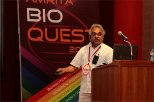
 Krishnakumar Menon, Ph.D.
Associate Professor, Centre for Nanosciences & Molecular Medicine, Amrita University, Kochi, India
A Far-Western Clinical Proteomics Approach to Detect Molecules of Clinical and Pathological Significance in the Neurodegenerative Disease Multiple Sclerosis.
Multiple Sclerosis (MS), an autoimmune neurodegenerative disorder of the central nervous system. The disease affects young adults at their prime age leading to severe debilitation over several years. Despite advances in MS research, the cause of the disease remains elusive. Thus, our objective is to identify novel molecules of pathological and diagnostic significance important in the understanding, early diagnosis and treatment of MS. Biological fluids such as cerebrospinal fluid (CSF), that bathe the brain serve as a potential source for the identification of pathologically significant autoantibody reactivity in MS. In this regard, we report the development of an unbiased clinical proteomics approach for the detection of reactive CSF molecules that target brain proteins from patients with MS. Proteins of myelin and myelin-axolemmal complexes were separated by two-dimensional gel electrophoresis, blotted onto membranes and probed separately with biotinylated unprocessed CSF samples. Protein spots that reacted specifically to MS-CSF’s were further analyzed by matrix assisted laser desorption ionization-time-of-flight time-of-flight mass spectrometry. In addition to previously reported proteins found in MS, we have identified several additional molecules involved in mitochondrial and energy metabolism, myelin gene expression and/or cytoskeletal organization. Among these identified molecules, the cellular expression pattern of collapsin response mediator protein-2 and ubiquitin carboxy-terminal hydrolase L1 were investigated in human chronic-active MS lesions by immunohistochemistry. The observation that in multiple sclerosis lesions phosphorylated collapsin response mediator protein-2 was increased, whereas Ubiquitin carboxy-terminal hydrolase L1 was down-regulated, not only highlights the importance of these molecules in the pathology of this disease, but also illustrates the use of our approach in attempting to decipher the complex pathological processes leading to multiple sclerosis and other neurodegenerative diseases. Further, we show that in clinicaly isolated syndrome (CIS), we could identify important molecules that could serve as an early diagnostic biomarker in MS.
 K. P. Mohanakumar, Ph.D.
Chief Scientist, Cell Biology & Physiology Division, Indian Institute of Chemical Biology, Kolkata
Neuroprotective and neurodestructive effects of Ayurvedic drug constituents: Parkinson’s disease
The present study reports the good and the bad entities in an Indian traditional medicine used for treating Parkinson’s disease (PD). A prospective clinical trial on the effectiveness of Ayurvedic medication in a population of PD patients revealed significant benefits, which has been attributed to L-DOPA present in the herbs [1]. Later studies revealed better benefits with one of the herbs alone, compared to pure L-DOPA in a clinical trial conducted in UK [2], and in several studies conducted on animal models of PD in independent laboratories world over [3-5]. We have adapted strategies to segregate molecules from the herb, and then carefully removed L-DOPA contained therein, and tested each of these sub-fractions for anti-PD activity in 1-methyl-4-phenyl-1,2,3,6-tetrahydropyridine, rotenone and 6-hydroxydopamine -induced parkinsonian animal models, and transgenic mitochondrial cybrids. We report here two classes of molecules contained in the herb, one of which possessed severe pro-parkinsonian (phenolic amine derivatives) and the other having excellent anti-parkinsonian potential (substituted tetrahydroisoquinoline derivatives). The former has been shown to cause severe dopamine depletion in the striatum of rodents, when administered acutely or chronically. It also caused significant behavioral aberrations, leading to anxiety and depression [6]. The latter class of molecules administered in PD animal model [7], caused reversal of behavioral dysfunctions and significant attenuation of striatal dopamine loss. These effects were comparable or better than the effects of the anti-PD drugs, selegiline or L-DOPA. The mechanism of action of the molecule has been found to be novel, at the postsynaptic receptor signaling level, as well as cellular α-synuclein oligomerization and specifically at mitochondria. The molecule helped in maintaining mitochondrial ETC complex activity and stabilized cellular respiration, and mitochondrial fusion-fission machinery with specific effect on the dynamin related protein 1. Although there existed significant medical benefits that could be derived to patients due to the synergistic actions of several molecules present in a traditional preparation, accumulated data in our hands suggest complicated mechanisms of actions of Ayurvedic medication. Our results also provide great hope for extracting, synthesizing and optimizing the activity of anti-parkinsonian molecules present in traditional Ayurvedic herbs, and for designing novel drugs with novel mechanisms of action.
- N, Nagashayana, P Sankarankutty, MRV Nampoothiri, PK Mohan and KP Mohanakumar, J Neurol Sci. 176, 124-7, 2000.
- Katzenschlager R, Evans A, Manson A, Patsalos PN, Ratnaraj N, Watt H, Timmermann L, Van der Giessen R, Lees AJ. J Neurol Neurosurg Psychiatry.75, 1672-7, 2004.
- Manyam BV, Dhanasekaran M, Hare TA. Phytother Res. 18, 706-12, 2004.
- Kasture S, Pontis S, Pinna A, Schintu N, Spina L, Longoni R, Simola N, Ballero M, Morelli M. Neurotox Res. 15, 111-22, 2009.
- Lieu CA, Kunselman AR, Manyam BV, Venkiteswaran K, Subramanian T. Parkinsonism Relat Disord.16, 458-65, 2010.
- T Sengupta and KP Mohanakumar, Neurochem Int. 57, 637-46, 2010.
- T Sengupta, J Vinayagam, N Nagashayana, B Gowda, P Jaisankar and KP Mohanakumar, Neurochem Res 36, 177-86, 2011
 Andrey Panteleyev, Ph.D.
Vice Chair, Division of Molecular Biology, NBICS Centre-Kurchatov Institute, Moscow, Russia
The system of PAS proteins (HIF and AhR) as an interface between environment and skin homeostasis
Regulation of normal skin functions as well as etiology of many skin diseases are both tightly linked to the environmental impact. Nevertheless, molecular aspects of skin-environment communication and mechanisms coordinating skin response to a plurality of environmental stressors remain poorly understood.
Our studies along with the work of other groups have identified the family of PAS dimeric transcription factors as an essential sensory and regulatory component of communication between skin and the environment. This protein family comprises a number of hypoxia-induced factors (HIF-alpha proteins), aryl hydrocarbon receptor (AhR), AhR nuclear translocator (ARNT), and several proteins implicated in control of rhythmic processes (Clock, Period, and Bmal proteins). Together, various PAS proteins (and first of all ARNT – as the central dimerization partner in the family) control such pivotal aspects of cell physiology as drug/xenobiotic metabolism, hypoxic and UV light response, ROS activity, pathogen defense, overall energy balance and breathing pathways.
In his presentation Dr. Panteleyev will focus on the role of ARNT activity and local hypoxia in control of keratinocyte differentiation and cornification. His recent work revealed that ARNT negatively regulates expression of late differentiation genes through modulation of amphiregulin expression and downstream alterations in activity of EGFR pathway. All these effects are highly dependent on epigenetic mechanisms such as histone deacetylation. Characterisation of hypoxia as a key microenvironmental factor in the skin and the role of HIF pathway in control of dermal vasculature and epidermal functions is another major focus of Dr. Panteleyev’s presentation.
In general, the studies of Dr. Panteleyev’s laboratory provide an insight into the PAS-dependent maintenance of skin homeostasis and point to the potential role of these proteins in pathogenesis of environmentally-modulated skin diseases such as barrier defects, desquamation abnormalities, psoriasis, etc.


