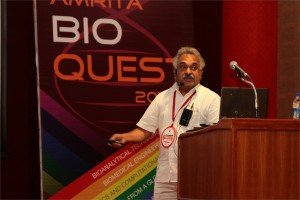
 Rohit Manchanda, Ph.D.
Professor, Biomedical Engineering Group, IIT-Bombay, India
Modelling the syncytial organization and neural control of smooth muscle: insights into autonomic physiology and pharmacology
We have been studying computationally the syncytial organization and neural control of smooth muscle in order to help explain certain puzzling findings thrown up by experimental work. This relates in particular to electrical signals generated in smooth muscles, such as synaptic potentials and spikes, and how these are explicable only if three-dimensional syncytial biophysics are taken fully into account. In this talk, I shall provide an illustration of outcomes and insights gleaned from such an approach. I shall first describe our work on the mammalian vas deferens, in which an analysis of the effects of syncytial coupling led us to conclude that the experimental effects of a presumptive gap junction uncoupler, heptanol, on synaptic potentials were incompatible with gap junctional block and could best be explained by a heptanol-induced inhibition of neurotransmitter release, thus compelling a reinterpretation of the mechanism of action of this agent. I shall outline the various lines of evidence, based on indices of syncytial function, that we adduced in order to reach this conclusion. We have now moved on to our current focus on urinary bladder biophysics, where the questions we aim to address are to do with mechanisms of spike generation. Smooth muscle cells in the bladder exhibit spontaneous spiking and spikes occur in a variety of distinct shapes, making their generation problematic to explain. We believe that the variety in shapes may owe less to intrinsic differences in spike mechanism (i.e., in the complement of ion channels participating in spike production) and more to features imposed by syncytial biophysics. We focus especially on the modulation of spike shape in a 3-D coupled network by such factors as innervation pattern, propagation in a syncytium, electrically finite bundles within and between which the spikes spread, and some degree of pacemaker activity by a sub-population of the cells. I shall report two streams of work that we have done, and the tentative conclusions these have enabled us to reach: (a) using the NEURON environment, to construct the smooth muscle syncytium and endow it with synaptic drive, and (b) using signal-processing approaches, towards sorting and classifying the experimentally recorded spikes.
 Gillian Murphy, Ph.D.
Professor, Department of Oncology, University of Cambridge, UK
A novel strategy for targeting metalloproteinases in cancer
Epithelial tumours evolve in a multi-step manner, involving both inflammatory and mesenchymal cells. Although intrinsic factors drive malignant progression, the influence of the micro-environment of neoplastic cells is a major feature of tumorigenesis. Extracellular proteinases, notably the metalloproteinases, are key players in the regulation of this cellular environment, acting as major effectors of both cell-cell and cell-extracellular matrix (ECM) interactions. They are involved in modifying ECM integrity, growth factor availability and the function of cell surface signalling systems, with consequent effects on cellular differentiation, proliferation and apoptosis.This has made metalloproteinases important targets for therapeutic interventions in cancer and small molecule inhibitors focussed on chelation of the active site zinc and binding within the immediate active site pocket were developed. These were not successful in early clinical trials due to the relative lack of specificity and precise knowledge of the target proteinase(s) in specific cancers. We can now appreciate that it is essential that we understand the relative roles of the different enzymes (of which there are over 60) in terms of their pro and anti tumour activity and their precise sites of expression The next generations of metalloproteinase inhibitors need the added specificity that might be gained from an understanding of the structure of individual active sites and the role of extra catalytic domains in substrate binding and other aspects of their biology. We have prepared scFv antibodies to the extra catalytic domains of two membrane metalloproteinases, MMP-14 and ADAM17, that play key roles in the tumour microenvironment. Our rationale and experiences with these agents will be presented in more detail.
 K. P. Mohanakumar, Ph.D.
Chief Scientist, Cell Biology & Physiology Division, Indian Institute of Chemical Biology, Kolkata
Neuroprotective and neurodestructive effects of Ayurvedic drug constituents: Parkinson’s disease
The present study reports the good and the bad entities in an Indian traditional medicine used for treating Parkinson’s disease (PD). A prospective clinical trial on the effectiveness of Ayurvedic medication in a population of PD patients revealed significant benefits, which has been attributed to L-DOPA present in the herbs [1]. Later studies revealed better benefits with one of the herbs alone, compared to pure L-DOPA in a clinical trial conducted in UK [2], and in several studies conducted on animal models of PD in independent laboratories world over [3-5]. We have adapted strategies to segregate molecules from the herb, and then carefully removed L-DOPA contained therein, and tested each of these sub-fractions for anti-PD activity in 1-methyl-4-phenyl-1,2,3,6-tetrahydropyridine, rotenone and 6-hydroxydopamine -induced parkinsonian animal models, and transgenic mitochondrial cybrids. We report here two classes of molecules contained in the herb, one of which possessed severe pro-parkinsonian (phenolic amine derivatives) and the other having excellent anti-parkinsonian potential (substituted tetrahydroisoquinoline derivatives). The former has been shown to cause severe dopamine depletion in the striatum of rodents, when administered acutely or chronically. It also caused significant behavioral aberrations, leading to anxiety and depression [6]. The latter class of molecules administered in PD animal model [7], caused reversal of behavioral dysfunctions and significant attenuation of striatal dopamine loss. These effects were comparable or better than the effects of the anti-PD drugs, selegiline or L-DOPA. The mechanism of action of the molecule has been found to be novel, at the postsynaptic receptor signaling level, as well as cellular α-synuclein oligomerization and specifically at mitochondria. The molecule helped in maintaining mitochondrial ETC complex activity and stabilized cellular respiration, and mitochondrial fusion-fission machinery with specific effect on the dynamin related protein 1. Although there existed significant medical benefits that could be derived to patients due to the synergistic actions of several molecules present in a traditional preparation, accumulated data in our hands suggest complicated mechanisms of actions of Ayurvedic medication. Our results also provide great hope for extracting, synthesizing and optimizing the activity of anti-parkinsonian molecules present in traditional Ayurvedic herbs, and for designing novel drugs with novel mechanisms of action.
- N, Nagashayana, P Sankarankutty, MRV Nampoothiri, PK Mohan and KP Mohanakumar, J Neurol Sci. 176, 124-7, 2000.
- Katzenschlager R, Evans A, Manson A, Patsalos PN, Ratnaraj N, Watt H, Timmermann L, Van der Giessen R, Lees AJ. J Neurol Neurosurg Psychiatry.75, 1672-7, 2004.
- Manyam BV, Dhanasekaran M, Hare TA. Phytother Res. 18, 706-12, 2004.
- Kasture S, Pontis S, Pinna A, Schintu N, Spina L, Longoni R, Simola N, Ballero M, Morelli M. Neurotox Res. 15, 111-22, 2009.
- Lieu CA, Kunselman AR, Manyam BV, Venkiteswaran K, Subramanian T. Parkinsonism Relat Disord.16, 458-65, 2010.
- T Sengupta and KP Mohanakumar, Neurochem Int. 57, 637-46, 2010.
- T Sengupta, J Vinayagam, N Nagashayana, B Gowda, P Jaisankar and KP Mohanakumar, Neurochem Res 36, 177-86, 2011
 Nader Pourmand, Ph.D.
Director, UCSC Genome Technology Center,University of California, Santa Cruz
Biosensor and Single Cell Manipulation using Nanopipettes
Approaching sub-cellular biological problems from an engineering perspective begs for the incorporation of electronic readouts. With their high sensitivity and low invasiveness, nanotechnology-based tools hold great promise for biochemical sensing and single-cell manipulation. During my talk I will discuss the incorporation of electrical measurements into nanopipette technology and present results showing the rapid and reversible response of these subcellular sensors to different analytes such as antigens, ions and carbohydrates. In addition, I will present the development of a single-cell manipulation platform that uses a nanopipette in a scanning ion-conductive microscopy technique. We use this newly developed technology to position the nanopipette with nanoscale precision, and to inject and/or aspirate a minute amount of material to and from individual cells or organelle without comprising cell viability. Furthermore, if time permits, I will show our strategy for a new, single-cell DNA/ RNA sequencing technology that will potentially use nanopipette technology to analyze the minute amount of aspirated cellular material.


