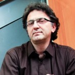
 Ayyappan Nair, Ph.D.
Head, Business Development (Technologies, Discovery Biology), Anthem Biosciences & DavosPharma, New Jersey, USA
Inhibition of NF-κB regulated gene expression by chrysoeriol suppresses tumorigenesis in breast cancer cells
Amrutha K1, Pandurangan Nanjan1, Sanu K Shaji1, Damu Sunilkumar1, Subhalakshmi K1, Rashmi U Nair1, Lakshmi Rajakrishna2, Asoke Banerji1, Ayyappan Ramesh Nair1*,2
- School of Biotechnology, Amrita Vishwa Vidyapeetham, Amritapuri Campus, Clappana P.O., Kollam – 690 525, Kerala, India
- Anthem Biosciences, No 49, Canara Bank Road, Bommasandra Industrial Area, Phase 1, Hosur Road, Bangalore – 560 099, Karnataka, India
Abstract: A large number of effective cancer-preventing compounds inhibit the activation of nuclear factor-κ B (NF-κB). It has been previously demonstrated that some flavonoids that are a vital component of our diet inhibits this pathway. As a consequence, many flavonoids inhibit genes involved in various aspects of tumorigenesis and have thus emerged as potential chemopreventive candidates for cancer treatment. We studied the effect of 17 different flavonoids, including the highly evaluated quercetin on the NF-κB pathway, and on the expression of MMP-9 and COX-2 (two NF-κB regulated genes involved in metastasis) in the highly invasive human breast cancer cell line MDA-MB-231. The findings suggest that not all the quercetin like flavone backbone compounds inhibit the NF-κB pathway, and that the highly hydoxylated flavonols quercetagetin and gossypetin did not inhibit this pathway, nor did it inhibit the expression of MMP-9 and COX-2. This indicates a correlation between inhibition of NF-κB and subsequent suppression of these NF-κB regulated genes. Here, we also report the novel observation that the not so well characterized methoxylated flavone chrysoeriol inhibited the NF-κB pathway, and was most potent in reducing the expression of MMP-9 and COX-2. Based on these observations, the cellular effects of chrysoeriol were evaluated in MDA-MB-231. Chrysoeriol caused cell cycle arrest at G2/M, inhibited migration and invasion, and caused cell death of macrophages that contributed to migration of these cancer cells. These effects of chrysoeriol make it a potential therapeutic candidate for breast cancer metastasis.

Binu K Aa, Jem Prabhakarb, Thara Sc and Lakshmi Sd,∗
aDepartment of Clinical Diagnostics Services and Translational Research, Malabar Cancer Centre, Thalassery, Kerala, India.
bDivision of Surgical Oncology, Division of Pathology
dDivision of Cancer Research, Regional Cancer Centre, Thiruvananthapuram, Kerala, India.
Introduction
AIB1, a member of the nuclear co activators, promotes the transcriptional activity of multiple nuclear receptors such as the ER and other transcription factors. Chemokines produced by stromal cells have potential to influence ERα-positive breast cancer progression to metastasis. CXCR4 is the physiological receptor for SDF1, together shown to stimulate the chemotactic and invasive behavior of breast cancer cells to serve as a homing mechanism to sites of metastasis. We propose that over expression of AIB1 in breast cancer cells leads to increased SDF1 and CXCR4 expression, which induces invasion and metastasis of cancer cells.
Materials and Methods
Breast tumor and normal breast tissues from patients in Regional Cancer Centre, Thiruvananthapuram were used for study. The modulatory effect of AIB1 was studied in MCF-7 cells with AIB1 siRNA transfection along with treatment of 17β-Estradiol (E2), 4-hydroxytamoxifen (4OHT), combinations of E2 and 4OHT. The gene expression pattern and protein localization were assessed by RT-PCR and immunofluorescence microscopy respectively. The metastatic and invasive properties were assessed by wound healing assay. Quantitative colocalization analyses were done to assess the association of proteins using Pearson’s correlation coefficient.
Result and Conclusion
The mRNA and protein level expression of AIB1, CXCR4 and SDF1 were higher in tumor samples than in normal samples. AIB1 was localized to the nuclei whereas CXCR4 and SDF1 immunoreactivity were observed in the cytoplasm and to a lesser extent in the nuclei of tumor epithelial cells. In tumor samples the gene level expressions of AIB1 showed significant positive correlations with SDF1(r = 0.213, p = 0.018). CXCR4 showed significant positive correlation with SDF1 in gene (r = 0.498, p = 0.000) and protein levels(r = 0.375, p = 0.002). Quantitative colocalization analyses showed a marked reduction in expression of CXCR4 and SDF1 in siAIB1MCF-7 cells than MCF-7 cells with different treatment groups. Wound healing assay shows reduced wound healing in siAIB1 treated MCF-7 cells.
In recent years, targeting specific cancer pathways and key molecules to arrest tumor growth and achieve tumor eradication have proven a challenge; due to acquired resistance and homing of cancer cells to various metastatic sites. The present study revealed that silencing AIB1 can prevent the over expression of SDF1 and CXCR4. Co activator levels determine the basal and estrogen-inducible expression of SDF1, a secreted protein that controls breast cancer cell proliferation and invasion through autocrine and paracrine mechanisms (Hall et al. 2003). The effects of CXCR4 overexpression has been correlated with SDF1 mediated activation of downstream signaling via ERK1/2 and p38 MAPK and with an enhancement of ER-mediated gene expression (Rhodes et al. 2011). It is possible that over expression of AIB1 as a stimulant involved in the expression of CXCR4 might up-regulate the expression of prometastatic and angiogenic genes. Thus based on these observations it can be concluded that SDF1/CXCR4 overexpression, with significant association with AIB1 expression, itself contribute to the development of mammary cancer and metastatic progression.
 Leland H. Hartwell Ph.D.
2001 Nobel Laureate, Physiology & Medicine
Dr. Lee Hartwell received the 2001 Nobel Prize in Physiology / Medicine for his discovery of protein molecules that control the division of cells. He was the President and Director of the Fred Hutchinson Cancer Research Center in Seattle, Washington before moving to Arizona State University’s Center for Sustainable Health.
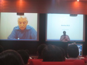
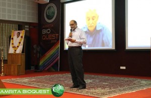
Dr. Hartwell is also adjunct faculty at Amrita University. He spoke to the delegates at Bioquest from his office in the US, over Amrita’s e-learning platform A-View. Given below are excerpts from his address.
I would like to address the young people in the audience. I know that many of you may have come to this meeting wondering, “How can I become a successful scientist? How can I prepare myself to make a contribution in this world?”
These questions are interesting to me also.
Believe it or not, I am still trying to be a successful scientist. That may surprise you since you probably think that a Nobel laureate must have found the answers. But the problem is that the answers to these questions change with time and the answers are different today than what they were when I began my career fifty years ago. The strategy of the 1960’s doesn’t work so well anymore. What is different now?
First, what we know now is much more. For example, by 1970, no genes from any organisms were sequenced. In 2013, we have the complete sequence of the human genome. Second, not only do we know much more today, accessing that knowledge is easy. Third, obtaining new information is much faster today.
Our rich understanding of science and technology is now needed to solve many serious problems. The human population has reached the size where we are utilizing all available resource of the planet. We are utilizing all of the agricultural land, all of the water, all of the forest and fishing resources. We are also polluting the planet that we live on.
We are polluting the land with fertilizers and pesticides; the oceans with acids and the atmosphere with carbon dioxide. We are using up top soil and ground water, thereby reducing our capacity to feed ourselves. We are using up petroleum, the energy source that our entire economy is dependent on. These are problems we were largely unaware of, fifty years ago. But these are problems that must be solved in your life times.
The big question facing your generation is, how can human beings live sustainably on planet earth. Your two broad goals on sustainability are 1) leave the planet as you first found it for your future generations; don’t use up the resources and don’t pollute the planet 2) everyone deserves to have an equal share of the earth’s resources.
Income strongly determines one’s opportunities in life. Many poor people succumb to chronic diseases and unhealthy environments. This inequality undermines our ability to live sustainably. We can’t ask the poor to leave the planet as they found it if they can’t support their families. Education, healthcare, employment are essential to having a sustainable society.
How can we be a successful scientist in 2013?
1. First choose a problem to solve
2. Ask questions to understand why it is not solved
3. Collaborate with those who can help
4. Develop a solution that works in the real worldChronic diseases are our major burden and this burden will get worse. Heart disease, diabetes, cancer, dementia and other diseases. The good news is that the chronic diseases are largely preventable and more easily curable if detected early. One question that attracts me is how can we detect disease earlier when it can be more easily cured?
Can we use our increasing knowledge in molecular biology to identify biomarkers for early disease detection?
We need to collaborate very closely with clinicians who care for patients to find out exactly where they need help.
I think if we apply our technology to important clinical questions we will actually save medical expenditure and be well on our way to making a great contribution to society.
 Nader Pourmand, Ph.D.
Director, UCSC Genome Technology Center,University of California, Santa Cruz
Biosensor and Single Cell Manipulation using Nanopipettes
Approaching sub-cellular biological problems from an engineering perspective begs for the incorporation of electronic readouts. With their high sensitivity and low invasiveness, nanotechnology-based tools hold great promise for biochemical sensing and single-cell manipulation. During my talk I will discuss the incorporation of electrical measurements into nanopipette technology and present results showing the rapid and reversible response of these subcellular sensors to different analytes such as antigens, ions and carbohydrates. In addition, I will present the development of a single-cell manipulation platform that uses a nanopipette in a scanning ion-conductive microscopy technique. We use this newly developed technology to position the nanopipette with nanoscale precision, and to inject and/or aspirate a minute amount of material to and from individual cells or organelle without comprising cell viability. Furthermore, if time permits, I will show our strategy for a new, single-cell DNA/ RNA sequencing technology that will potentially use nanopipette technology to analyze the minute amount of aspirated cellular material.
 Satheesh Babu T. G., Ph.D.
Associate Professor, Department of Sciences, School of Engineering, Amrita University, Coimbatore, India
Nanomaterials for ‘enzyme-free’ biosensing
Enzyme based sensors have many draw backs such as poor storage stability, easily affected by the change in pH and temperature and involves complicated enzyme immobilization procedures. To address this limitation, an alternative approach without the use of enzyme, “non-enzymatic” has been tried recently. Choosing the right catalyst for direct electrochemical oxidation / reduction of a target molecule is the key step in the fabrication of non-enzymatic sensors.
Non-enzymatic sensors for glucose, creatinine, vitamins and cholesterol are fabricated using different nanomaterials, such as nanotubes, nanowires and nanoparticles of copper oxide, titanium dioxide, tantalum oxide, platinum, gold and graphenes. These sensors selectively catalyse the targeted analyte with very high sensitivity. These nanomaterials based sensors combat the drawbacks of enzymatic sensors.

Anupama Natarajan, James Hickman and Peter Molnar
Novel Cell-Based Biosensors for High Throughput Toxin Detection and Drug Screening Applications
Over the last decade there has been focus on the development of cellbased biosensors to detect environmental toxins or to combat the threats of biological warfare. These sensors have been shown to have multiple applications including understanding function and behaviour at the cellular and tissue levels, in cell electrophysiology and as drug screening tools that can eliminate animal testing. These factors make the development of cell-based biosensors into high throughput systems a priority in pharmacological, environmental and defence industries (Pancrazio J J et al. 1999, Kang G et al. 2009, Krinke D et al. 2009). We have developed a high through-put in vitro cell-silicon hybrid platform that could be used to analyze both cell function and response to various toxins and drugs. Our hypothesis was that by utilizing surface modification to provide external guidance cues as well as optimal growth conditions for different cell types (Cardiac and Neuronal), we could enhance the information output and content of such a system. An intrinsic part of this study was to create ordered or patterned functional networks of cells on Micro-electrode arrays (MEA). Such engineered networks had a two-fold purpose in that they not only aided in a more accurate analysis of cell response and cell and tissue behaviour, but also increased the efficiency of the system by increasing the connectivity and placement of the cells over the recording electrodes. Here we show the response of this system to various toxins and drugs and the measurement of several vital cardiac parameters like conduction velocity and refractory period (Natarajan A et al. 2011)
 Jeff Perry, Ph.D.
Assistant Professor, University of California, Riverside
Combined Crystallography and SAXS Methods for Studying Macromolecular Complexes
Recent developments in small angle X-ray scattering (SAXS) are rapidly providing new insights into protein interactions, complexes and conformational states in solution, allowing for detailed biophysical quantification of samples of interest1. Initial analyses provide a judgment of sample quality, revealing the potential presence of aggregation, the overall extent of folding or disorder, the radius of gyration, maximum particle dimensions and oligomerization state. Structural characterizations may include ab initio approaches from SAXS data alone, or enhance structural solutions when combined with previously determined crystal/NMR domains. This combination can provide definitions of architectures, spatial organizations of the protein domains within a complex, including those not yet determined by crystallography or NMR, as well as defining key conformational states. Advantageously, SAXS is not generally constrained by macromolecule size, and rapid collection of data in a 96-well plate format provides methods to screen sample conditions. Such screens include co-factors, substrates, differing protein or nucleotide partners or small molecule inhibitors, to more fully characterize the variations within assembly states and key conformational changes. These analyses are also useful for screening constructs and conditions that are most likely to promote crystal growth. Moreover, these high throughput structural determinations can be leveraged to define how polymorphisms affect assembly formations and activities. Also, SAXS-based technologies may be potentially used for novel structure-based screening, for compounds inducing shape changes or associations/diassociations. This is addition to defining architectural characterizations of complexes and interactions for systems biology-based research, and distinctions in assemblies and interactions in comparative genomics. Thus, SAXS combined with crystallography/NMR and computation provides a unique set of tools that should be considered as being part of one’s repertoire of biophysical analyses, when conducting characterizations of protein and other macromolecular interactions.
1 Perry JJ & Tainer JA. Developing advanced X-ray scattering methods combined with crystallography and computation. Methods. 2013 Mar;59(3):363-71.


