


 Shyam Diwakar, Ph.D.
Assistant Professor, Amrita School of Biotechnology
 Jaap Heringa, Ph.D.
Director & Professor of Bioinformatics, IBIVU VU University Amsterdam, The Netherlands
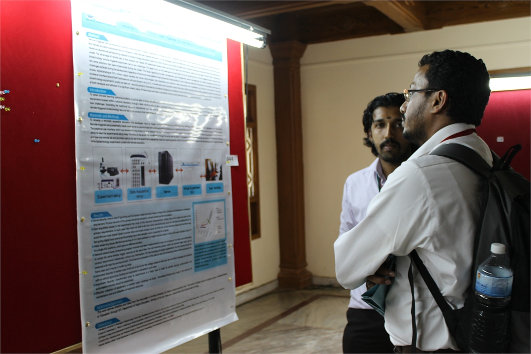
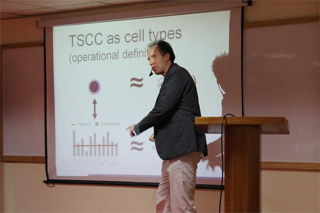
Modeling strategy based on Petri-nets
In my talk I will introduce a formal modeling strategy based on Petri-nets, which are a convenient means of modeling biological processes. I will illustrate the capabilities of Petri-nets as reasoning vehicles using two examples: Haematopoietic stem cell differentiation in mice, and vulval development in C. elegance. The first system was modeled using a Boolean implementation, and the second using a coarse-grained multi-cellular Petri-net model. Concepts such as the model state space, attractor states, and reasoning to adapt the model to the biological reality will be discussed.
 Rajgopal Srinivasan, Ph.D.
Principal Scientist & Head Bio IT R&D, TCS Innovation Labs, India
Interpretation of Genomic Variation – Identifying Rare Variations Leading to Disease
Genome sequencing technologies are generating an abundance of data on human genetic variations. A big challenge lies in interpreting the functional relevance of such variations, especially in clinical settings. A first step in understanding the clinical relevance of genetic variations is to annotate the variants for region of occurrence, degree of conservation both within and across species, pattern of variation across related individuals, novelty of the variation and know effects of related variations. Several tools already exist for this purpose. However, these tools have their strengths and weaknesses. A second issue is the development of algorithms, which, given a rich annotation of variants are able to prioritize the variants as being relevant to the phenotype under investigation.
In my talk I will detail work that has been done in our labs to address both of the above problems. I will also illustrate the application of these tools that helped identify a rare mutation in the ATM gene leading to a diagnosis of AT in two infants.
 Ajay Shah, Ph.D.
 Kal Ramnarayan, Ph.D.
Co-founder President & Chief Scientific Officer, Sapient Discovery, San Diego, CA, USA
A cost-effective approach to Protein Structure-guided Drug Discovery: Aided by Bioinformatics, Chemoinformatics and computational chemistry
With the mapping of the human genome completed almost a decade ago, efforts are still underway to understand the gene products (i.e., proteins) in the human biological and disease pathways. Deciphering such information is very important for the discovery and development of small molecule drugs as well as protein therapeutics for various human diseases for which no cure exists. As an example, with more than 500 members, the kinase family of protein targets continues to be an important and attractive class for drug discovery. While how many of the members in this family are actually druggable is still to be established, there are several ongoing efforts on this class of proteins across a broad spectrum of disease categories. Even though in general the protein structural topology might looks similar, there are issues with respect selectivity of identified small molecule inhibitors when, the lead molecule discovery is carried out at the ATP binding site. As an added complexity, allosteric modulators are needed for some of the members, but the actual site for such modulation on the protein target can not resolved with uncertainty. In this presentation we will describe a bioinformatics and computational based platform for small molecule discovery for protein targets that are involved in protein-protein interactions as well as targets like kinases and phosphatases. We will describe a computational approach in which we have used an informatics based platform with several hundred kinases to sort through in silico and identify inhibitors that are likely to be highly selective in the lead generation phase. We will discuss the implication of this approach on the drug discovery of the kinase and phosphatase classes in general and independent of the disease category.
 Prashanth Athri, Ph.D.
Senior Specialist, Strand Life Sciences, Bengaluru, India
Rare disease diagnostic platform
At Strand, genomic sequencing combined with bioinformatic analysis have provided discriminative diagnosis in the case of rare genetic disorders. Inspired by these cases, we are building an integrated software that combines curated literature content and bioinformatics databases with a clinically oriented user interface to substantially compress time taken to determine likely candidate genetic variants in a Diagnostic Odyssey. At the back end we employ various algorithms that systematically query our diverse knowledgebase to provide the clinicians a comprehensive, and possibly multidimensional, annotation of the variant in the context of disease.
 Karmeshu, Ph.D.
Dean & Professor, School of Computer & Systems Sciences & School of Computational & Integrative Sciences, Jawaharlal Nehru University, India.
Interspike Interval Distribution of Neuronal Model with distributed delay: Emergence of unimodal, bimodal and Power law
The study of interspike interval distribution of spiking neurons is a key issue in the field of computational neuroscience. A wide range of spiking patterns display unimodal, bimodal ISI patterns including power law behavior. A challenging problem is to understand the biophysical mechanism which can generate the empirically observed patterns. A neuronal model with distributed delay (NMDD) is proposed and is formulated as an integro-stochastic differential equation which corresponds to a non-markovian process. The widely studied IF and LIF models become special cases of this model. The NMDD brings out some interesting features when excitatory rates are close to inhibitory rates rendering the drift close to zero. It is interesting that NMDD model with gamma type memory kernel can also account for bimodal ISI pattern. The mean delay of the memory kernels plays a significant role in bringing out the transition from unimodal to bimodal ISI distribution. It is interesting to note that when a collection of neurons group together and fire together, the ISI distribution exhibits power law.
 Lalitha Subramanian, Ph.D.
Chief Scientific Officer & VP, Services at Scienomics, USA
Nanoscale Simulations – Tackling Form and Formulation Challenges in Drug Development and Drug Delivery
Lalitha Subramanian, Dora Spyriouni, Andreas Bick, Sabine Schweizer, and Xenophon Krokidis Scienomics
The discovery of a compound which is potent in activity against a target is a major milestone in Pharmaceutical and Biotech industry. However, a potent compound is only effective as a therapeutic agent when it can be administered such that the optimal quantity is transported to the site of action at an optimal rate. The active pharmaceutical ingredient (API) has to be tested for its physicochemical properties before the appropriate dosage form and formulation can be designed. Some of the commonly evaluated parameters are crystal forms and polymorphs, solubility, dissolution behavior, stability, partition coefficient, water sorption behavior, surface properties, particle size and shape, etc. Pharmaceutical development teams face the challenge of quickly and efficiently determining a number of properties with small quantities of the expensive candidate compounds. Recently the trend has been to screen these properties as early as possible and often the candidate compounds are not available in sufficient quantities. Increasingly, these teams are leveraging nanoscale simulations similar to those employed by drug discovery teams for several decades. Nanoscale simulations are used to predict the behavior using very little experimental data and only if this is promising further experiments are done. Another aspect where nanoscale simulations are being used in drug development and drug delivery is to get insights into the behavior of the system so that process failures can be remediated and formulation performance can be improved. Thus, the predictive screening and the in-depth understanding leads to experimental efficiency resulting in far-reaching business impacts.
With specific examples, this talk will focus on the different types of nanoscale simulations used to predict properties of the API in excipients and also provide insight into system behavior as a function of shelf life, temperature, mechanical stress, etc.
 Srisairam Achuthan, Ph.D.
Senior Scientific Programmer, Research Informatics Division, Department of Information Sciences, City of Hope, CA, USA
Applying Machine learning for Automated Identification of Patient Cohorts
Srisairam Achuthan, Mike Chang, Ajay Shah, Joyce Niland
Patient cohorts for a clinical study are typically identified based on specific selection criteria. In most cases considerable time and effort are spent in finding the most relevant criteria that could potentially lead to a successful study. For complex diseases, this process can be more difficult and error prone since relevant features may not be easily identifiable. Additionally, the information captured in clinical notes is in non-coded text format. Our goal is to discover patterns within the coded and non-coded fields and thereby reveal complex relationships between clinical characteristics across different patients that would be difficult to accomplish manually. Towards this, we have applied machine learning techniques such as artificial neural networks and decision trees to determine patients sharing similar characteristics from available medical records. For this proof of concept study, we used coded and non-coded (i.e., clinical notes) patient data from a clinical database. Coded clinical information such as diagnoses, labs, medications and demographics recorded within the database were pooled together with non-coded information from clinical notes including, smoking status, life style (active / inactive) status derived from clinical notes. The non-coded textual information was identified and interpreted using a Natural Language Processing (NLP) tool I2E from Linguamatics.

Kunal Kundu, Sushma Motamarri, Uma Sunderam, Steven E. Brenner and Rajgopal Srinivasan.
VARANT: The Variant Annotation Tool
Genome sequencing technologies are generating an abundance of data on human genetic variations. A big challenge lies in interpreting the functional relevance of such variations, especially in clinical settings. A first step in understanding the clinical relevance of genetic variations is to annotate the variants for region of occurrence, degree of conservation both within and across species, pattern of variation across related individuals, novelty of the variation and know effects of related variations. Several tools already exist for this purpose. However, these tools have their strengths and weaknesses. We will present an open-source tool, VARANT, written in the python programming language, that is easily extended to incorporate newer annotations.
A detailed variant annotation places variants in context, highlights significant findings and prioritizes candidates for further analysis. With this outlook we developed VARANT to annotate, prioritize and visualize variants. VARANT has 5 levels of annotation – genomic position based, gene based, untranslated region (UTR) based, mutation effect prediction and gene level disease association. The databases used for annotations have been compiled from several sources. The genomic position based annotation comprises of tagging variants present in dbSNP and 1000 Genomes projects, GWAS variants, variants in functionally constrained region and variants overlapping epigenetic signals. The gene-based annotation includes, the distance from splice sites for intronic variants; gene, transcript, amino acid change and splicing silencer and enhancers information for exonic variants. UTR based annotations comprise of UTR functional sites like miRNA binding site, internal ribosomal entry site, variations and deletions in UTR5-Coding Sequence(CDS) boundary, exon-intron boundary and CDS-UTR3 boundary.Mutation effect predictions are incorporated from PolyPhen2 and SIFT. Thus, a detailed annotation with VARANT captures multiple biological aspects of a variant and helps in filtering variants based on disease context. The input and output of VARANT is the universal Variant Call Format, with facilities to export the annotations to popular formats such as comma/tab separated values and MS Excel. Using a desktop computer with single core and 4GB RAM VARANT annotates over 50,000 variants/minute and can be readily parallelized. Being an exhaustive annotator with good performance using modest computational hardware, VARANT is a useful annotation tool for analyzing genomic variants. Furthermore, the tool includes facilities to update the underlying data sources in an automated fashion, and is easily extended to add additional annotations. VARANT also provides an interface to visualize variants in an annotated VCF file and to filter variants interactively based on annotation features like – region, mutation effect etc, and inheritance models. In addition to annotation, there are ongoing efforts to incorporate a variant prioritization module using the annotated features as well as inheritance information.
Bhadrachalam Chitturi, Balaji Raghavachari and Donghyun Kim
Efficient gene prioritization
The gene prioritization, GP, problem seeks to identify the most promising genes among several candidate genes. In genetics, gene related conditions are typically associated with chromosomal regions, say with GWAS. These associations yield lists of candidate genes. A priori, some genes i.e. seed genes, are associated with a specific disease D; additional genes that are implicated via associations constitute the potential candidates. Thus, most promising novel candidates for D are sought. In network based approach, a protein protein interaction network, i.e. NP , and a set S of seed genes constitute the prior knowledge. We treat a gene and the protein that it encodes identically. Various GP algorithms based on guilt by association are run on the NP to predict novel candidates [1–6]. They rank a new candidate gene by its estimated association to D.
Distance between a pair of genes is the shortest path measured in the number of edges. Diameter of a set of genes is the longest distance between any pair of genes in terms of the number of edges. The density of a set X of genes is defined as e(X)/|X| where e(X) denotes the number of edges among genes of X and |X| denotes the number of genes of X. The set S: (i) can be of minimal size (say one), (ii) is tightly coupled in NP , i.e. has low-diameter/high-density, or (iii) is loosely coupled, i.e. has high-diameter/low-density. Similarly, the GP algorithms can be partitioned into: Type-1 that ignore the edge weights and Type-2 that employ the edge weights. However, currently, the prioritization process neither exploits the character of S nor the type of GP algorithm that is run. Given S, we compute two core networks of NP which we call NC1 and NC2 that are subnetworks of NP . The idea is to execute GP algorithms of Type-1 and Type-2 on NC1 and NC2 respectively instead of NP . Typically, NC1 and NC2 are much smaller than NP . Also, one runs several algorithms of Type-1 and Type-2 [2–4, 6] and takes consensus [6].
In general, the time to run a GP algorithm say AP on NP i.e. t1 or to compute NC1 and NC2 i.e. t2 is proportional to e(NP ) where e(NP ) e(NC1) and e(NP ) e(NC2). However, executing AP on NC1/NC2 (a much smaller network) is much more efficient than executing AP on NP . We run several GP algorithms onNC1/NC2 [6] but computeNC1/NC2 only once. So, overall our method is more efficient. Preliminary implementation results show that for several GP algorithms, the candidates identified by our method match the topmost prioritized candidates identified by the direct execution of the algorithm on NP . Overall, our method was more efficient. Based on the number of candidates that we seek and the nature of S, we can generate variants of NCx, x ∈ {1, 2}. In some cases, AP determines the appropriate variant of NCx.
Nitish Sathyanrayanan, Sandesh Ganji and Holenarsipur Gundurao Nagendra.
Insilico Analysis of hypothetical proteins from Leishmania donovani: A Case study of a membrane protein of the MFS class reveals their plausible roles in drug resistance
Kala-azar or visceral leishmaniais (VL), caused by protozoan parasite Leishmania donovani, is one of the leading causes of morbidity and mortality in Bihar, India (Guerin et al. 2002; Mubayi et al. 2010). The disease is transmitted to the humans mainly by the vector, Phlebotmus argentipes, commonly known as Sand fly. The majority of VL (> 90%) occurs in only six countries: Bangladesh, India, Nepal, Sudan, Ethiopia and Brazil (Chappuis et al. 2007). In the Indian subcontinent, about 200 million people are estimated to be at risk of developing VL and this region harbors an estimated 67% of the global VL disease burden. The Bihar state only has captured almost 50% cases out of total cases in Indian sub-continent (Bhunia et al. 2013). ‘Conserved hypothetical’ proteins pose a challenge not just to functional genomics, but also to biology in general (Galperin and Koonin 2004). Leishmania donovani (strain BPK282A1) genome consists of a staggering ∼65% of hypothetical proteins. These uncharacterized proteins may enable better appreciation of signalling pathways, general metabolism, stress response and even drug resistance.
Rajasekhar Chekkara, Venkata Reddy Gorla and Sobha Rani Tenkayala
Pharmacophore modeling, atom-based 3D-QSAR and molecular docking studies on Pyrimido[5,4-e][1,2,4]triazine derivatives as PLK 1 inhibitors
Polo-like kinase 1 (PLK1) is a significant enzyme with diverse biological actions in cell cycle progression, specifically mitosis. Suppression of PLK1 activity by small molecule inhibitors has been shown to inhibit cancer, being BI 2536 one of the most potent active inhibitor of PLK1 mechanism. Pharmacophore modeling, atom-based 3D-QSAR and molecular docking studies were carried out for a set of 54 compounds belonging to Pyrimido[5,4-e][1,2,4]triazine derivatives as PLK1 inhibitors. A six-point pharmacophoremodel AAADDR, with three hydrogen bond acceptors (A), two hydrogen bond donors (D) and one aromatic ring (R) was developed by Phase module of Schrdinger suite Maestro 9. The generated pharmacophore model was used to derive a predictive atom-based 3D quantitative structure-activity relationship analysis (3D-QSAR) model for the training set (r2 = 0.88, SD = 0.21, F = 57.7, N = 44) and for test set (Q2 = 0.51, RMSE = 0.41, PearsonR = 0.79, N = 10). The original set of compounds were docked into the binding site of PLK1 using Glide and the active residues of the binding site were analyzed. The most active compound H18 interacted with active residues Leu 59, Cys133 (glide score = −10.07) and in comparison of BI 2536, which interacted with active residues Leu 59, Cys133 (glide score = −10.02). The 3D-QSAR model suggests that hydrophobic and electron-withdrawing groups are essential for PLK1 inhibitory activity. The docking results describes the hydrogen bond interactions with active residues of these compounds. These results which may support in the design and development of novel PLK1 inhibitors.
 Jeff Perry, Ph.D.
Assistant Professor, University of California, Riverside
Combined Crystallography and SAXS Methods for Studying Macromolecular Complexes
Recent developments in small angle X-ray scattering (SAXS) are rapidly providing new insights into protein interactions, complexes and conformational states in solution, allowing for detailed biophysical quantification of samples of interest1. Initial analyses provide a judgment of sample quality, revealing the potential presence of aggregation, the overall extent of folding or disorder, the radius of gyration, maximum particle dimensions and oligomerization state. Structural characterizations may include ab initio approaches from SAXS data alone, or enhance structural solutions when combined with previously determined crystal/NMR domains. This combination can provide definitions of architectures, spatial organizations of the protein domains within a complex, including those not yet determined by crystallography or NMR, as well as defining key conformational states. Advantageously, SAXS is not generally constrained by macromolecule size, and rapid collection of data in a 96-well plate format provides methods to screen sample conditions. Such screens include co-factors, substrates, differing protein or nucleotide partners or small molecule inhibitors, to more fully characterize the variations within assembly states and key conformational changes. These analyses are also useful for screening constructs and conditions that are most likely to promote crystal growth. Moreover, these high throughput structural determinations can be leveraged to define how polymorphisms affect assembly formations and activities. Also, SAXS-based technologies may be potentially used for novel structure-based screening, for compounds inducing shape changes or associations/diassociations. This is addition to defining architectural characterizations of complexes and interactions for systems biology-based research, and distinctions in assemblies and interactions in comparative genomics. Thus, SAXS combined with crystallography/NMR and computation provides a unique set of tools that should be considered as being part of one’s repertoire of biophysical analyses, when conducting characterizations of protein and other macromolecular interactions.
1 Perry JJ & Tainer JA. Developing advanced X-ray scattering methods combined with crystallography and computation. Methods. 2013 Mar;59(3):363-71.

 Akhilesh Pandey, Ph.D.
Professor, Johns Hopkins University School of Medicine, Baltimore, USA
A draft map of the human proteome
We have generated a draft map of the human proteome through a systematic and comprehensive analysis of normal human adult tissues, fetal tissues and hematopoietic cells as an India-US initiative. This unique dataset was generated from 30 histologically normal adult tissues, fetal tissues and purified primary hematopoietic cells that were analyzed at high resolution in the MS mode and by HCD fragmentation in the MS/MS mode on LTQ-Orbitrap Velos/Elite mass spectrometers. This dataset was searched against a 6-frame translation of the human genome and RNA-Seq transcripts in addition to standard protein databases. In addition to confirming a large majority (>16,000) of the annotated protein-coding genes in humans, we obtained novel information at multiple levels: novel protein-coding genes, unannotated exons, novel splice sites, proof of translation of pseudogenes (i.e. genes incorrectly annotated as pseudogenes), fused genes, SNPs encoded in proteins and novel N-termini to name a few. Many proteins identified in this study were identified by proteomic methods for the first time (e.g. hypothetical proteins or proteins annotated based solely on their chromosomal location). We have generated a catalog of proteins that show a more tissue-restricted pattern of expression, which should serve as the basis for pursuing biomarkers for diseases pertaining to specific organs. This study also provides one of the largest sets of proteotypic peptides for use in developing MRM assays for human proteins. Identification of several novel protein-coding regions in the human genome underscores the importance of systematic characterization of the human proteome and accurate annotation of protein-coding genes. This comprehensive dataset will complement other global HUPO initiatives using antibody-based as well as MRM mass spectrometry-based strategies. Finally, we believe that this dataset will become a reference set for use as a spectral library as well as for interesting interrogations pertaining to biomedical as well as bioinformatics questions.

 Tobias Preckel, Ph.D.
Life Science Center Manager, Agilent Technologies, Germany
Comprehensive characterization of Biopharmaceuticals: challenges and opportunities

Syed Salman Lateef and Vinayak A K
Development of Supercritical Fluid Chromatography methods for the replacement of existing USP Normal phase liquid chromatography methods
Normal phase liquid chromatography methods often have long run times and involve environmentally toxic/costly solvents. Supercritical chromatography methods on the other hand are faster, inexpensive, and eco-friendly. The low viscous supercritical carbon dioxide operates at high flow rates compared to LC without losing separation efficiency. In this work, SFC methods are developed to replace three United States Pharmacopeial (USP) normal phase achiral methods – prednisolone, tolazamide and cholecalciferol. System suitability parameters of the normal phase method are compared against the SFC method. Precision, linearity and robustness of the new SFC methods are demonstrated. SFC methods were found to be cost effective in terms of analysis time and solvent savings. The SFC method does not require purchase and disposal of expensive environmentally hazardous chemicals. Hence, the newly developed SFC method provides a faster and safer solution.
 Sudarslal S, Ph.D.
Associate Professor, School of Biotechnology, Amrita University
Electrospray ionization ion trap mass spectrometry for cyclic peptide characterization
There has been considerable interest in the isolation and structural characterization of bioactive peptides produced by bacteria and fungi. Most of the peptides are cyclic depsipeptides characterized by the presence of lactone linkages and β-hydroxy fatty acids. Occurrence of microheterogeneity is another remarkable property of these peptides. Even if tandem mass spectrometers are good analytical tools to structurally characterize peptides and proteins, sequence analysis of cyclic peptides is often ambiguous due to the random ring opening of the peptides and subsequent generation of a set of linear precursor ions with the same m/z. Here we report combined use of chemical derivatization and multistage fragmentation capability of ion trap mass spectrometers to determine primary sequences of a series of closely related cyclic peptides.


Ravindra Gudihal, Suresh Babu C V
Bioanalytical Characterization of Therapeutic Proteins
The characterization of therapeutic proteins such as monoclonal antibody (mAb) during different stages of manufacturing is crucial for timely and successful product release. Regulatory agencies require a variety of analytical technologies for comprehensive and efficient protein analysis. Electrophoresis-based techniques and liquid chromatography (LC) either standalone or coupled to mass spectrometry (MS) are at the forefront for the in-depth analysis of protein purity, isoforms, stability, aggregation, posttranslational modifications, PEGylation, etc. In this presentation, a combination of various chromatographic and electrophoretic techniques such as liquid-phase isoelectric focusing, microfluidic and capillary-based electrophoresis (CE), liquid chromatography (LC) and combinations of those with mass spectrometry techniques will be discussed. We present a workflow based approach to the analysis of therapeutic proteins. In successive steps critical parameters like purity, accurate mass, aggregation, peptide sequence, glycopeptide and glycan analysis are analyzed. In brief, the workflow involved proteolytic digestion of therapeutic protein for peptide mapping, N-Glycanase and chemical labeling reaction for glycan analysis, liquid-phase isoelectric focusing for enrichment of charge variants followed by a very detailed analysis using state of the art methods such as CE-MS and LC-MS. For the analysis of glycans, we use combinations of CE-MS and LC-MS to highlight the sweet spots of these techniques. CE-MS is found to be more useful in analysis of highly sialylated glycans (charged glycans) while nano LC-MS seems to be better adapted for analysis of neutral glycans. These two techniques can be used to get complementary data to profile all the glycans present in a given protein. In addition, microfluidic electrophoresis was used as a QC tool in initial screening for product purity, analysis of papain digestion fragments of mAb, protein PEGylation products, etc. The described workflow involves multiple platforms, provides an end to end solution for comprehensive protein characterization and aims at reducing the total product development time.
Tejaswini Subbannayya, Nandini A. Sahasrabuddhe, Arivusudar Marimuthu, Santosh Renuse, Gajanan Sathe, Srinivas M. Srikanth, Mustafa A. Barbhuiya, Bipin Nair, Juan Carlos Roa, Rafael Guerrero-Preston, H. C. Harsha, David Sidransky, Akhilesh Pandey, T. S. Keshava Prasad and Aditi Chatterjee
Proteomic profiling of gallbladder cancer secretome – a source for circulatory biomarker discovery
Gallbladder cancer (GBC) is the fifth most common cancer of the gastrointestinal tract and one of the common malignancies that occur in the biliary tract (Misra et al. 2006; Lazcano-Ponce et al. 2001). It has a poor prognosis with survival of less than 5 years in 90% of the cases (Misra et al. 2003). The etiology is ill-defined. Several risk factors have been reported including cholelithiasis, obesity, female gender and exposure to carcinogens (Eslick 2010; Kumar et al. 2006). Poor prognosis in GBC is mainly due to late presentation of the disease and lack of reliable biomarkers for early diagnosis. This emphasizes the need to identify and characterize cancer biomarkers to aid in the diagnosis and prognosis of GBC. Secreted proteins are an important class of molecules which can be detected in body fluids and has been targeted for biomarker discovery. There are challenges faced in the proteomic interrogation of body fluids especially plasma such as low abundance of tumor secreted proteins, high complexity and high abundance of other proteins that are not released by the tumor cells (Tonack et al. 2009). Profiling of conditioned media from the cancer cell lines can be used as an alternate means to identify secreted proteins from tumor cells (Kashyap et al. 2010; Marimuthu et al. 2012). We analyzed the invasive property of 7 GBC cell lines (SNU-308, G-415, GB-d1, TGBC2TKB, TGBC24TKB, OCUG-1 and NOZ). Four cell lines were selected for analysis of the cancer secretome based on the invasive property of the cells. We employed isobaric tags for relative and absolute quantitation (iTRAQ) labeling technology coupled with high resolution mass spectrometry to identify and characterize secretome from the panel of 4GBC cancer cells mentioned above. In total, we have identified around 2,000 proteins of which 175 were secreted at differential abundance across all the four cell lines. This secretome analysis will act as a reservoir of candidate biomarkers. Currently, we are investigating and validating these candidate markers from GBC cell secretome. Through this study, we have shown mass spectrometry-based quantitative proteomic analysis as a robust approach to investigate secreted proteins in cancer cells.