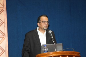
Vural Özdemir Ph.D.
Sanjeeva Srivastava Ph.D.
 Bharat B. Chattoo, Ph.D.
Professor, Faculty of Science M.S.University of Baroda, India
Biology of plant infection by Magnaporthe oryzae
The rice blast disease caused by the ascomycetous fungus Magnaporthe oryzae is a major constraint in rice production. Rice-M.oryzae is also emerging as a good model patho-system to investigate how the fungus invades and propagates within the host. Identification and characterisation of genes critical for fungal pathogenesis provides opportunities to explore their use as possible targets for development of strategies for combating fungal infection and to better understand the complex process of host-pathogen interaction.
We have used insertional mutagenesis and RNAi based approaches to identify pathogenesis related genes in this fungus. A large number of mutants were isolated using Agrobacterium tumefaciens mediated transformation (ATMT). Characterisation of several interesting mutants is in progress. We have identified a novel gene, MGA1, required for the development of appressoria. The mutant mga1 is unable to infect and is impaired in glycogen and lipid mobilization required for appressorium development. The glycerol content in the mycelia of the mutant was significantly lower as compared to wild type and it was unable to tolerate hyperosmotic stress. A novel ABC transporter was identified in this fungus. The abc4 mutant did not form functional appressoria, was non-pathogenic and showed increased sensitivity to certain antifungal molecules implying the role of ABC4 in multidrug resistance (MDR). Another mutant MoSUMO (MGG_05737) was isolated using a Split Marker technique; the mutant showed defects in growth, germination and infection. Immuno-fluorescence microscopy revealed that MoSumo is localized to septa in mycelia and nucleus as well as septa in spores. Two Dimensional Gel Electrophoresis showed differences in patterns of protein expression between Wild Type B157 and MoΔSumo mutant. We also isolated and charaterised mutants in MoALR2 (MGG_08843) and MoMNR2 (MGG_09884). Our results indicate that both MoALR2 and MoMNR2 are Mg2+ transporters, and the reduction in the levels of CorA transporters caused defects in surface hydrophobicity, cell wall stress tolerance, sporulation, appressorium formation and infection are mediated through changes in the key signaling cascades in the knock-down transformants. (Work supported by the Department of Biotechnology, Government of India)
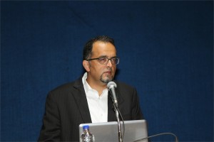
 Rohit Manchanda, Ph.D.
Professor, Biomedical Engineering Group, IIT-Bombay, India
Modelling the syncytial organization and neural control of smooth muscle: insights into autonomic physiology and pharmacology
We have been studying computationally the syncytial organization and neural control of smooth muscle in order to help explain certain puzzling findings thrown up by experimental work. This relates in particular to electrical signals generated in smooth muscles, such as synaptic potentials and spikes, and how these are explicable only if three-dimensional syncytial biophysics are taken fully into account. In this talk, I shall provide an illustration of outcomes and insights gleaned from such an approach. I shall first describe our work on the mammalian vas deferens, in which an analysis of the effects of syncytial coupling led us to conclude that the experimental effects of a presumptive gap junction uncoupler, heptanol, on synaptic potentials were incompatible with gap junctional block and could best be explained by a heptanol-induced inhibition of neurotransmitter release, thus compelling a reinterpretation of the mechanism of action of this agent. I shall outline the various lines of evidence, based on indices of syncytial function, that we adduced in order to reach this conclusion. We have now moved on to our current focus on urinary bladder biophysics, where the questions we aim to address are to do with mechanisms of spike generation. Smooth muscle cells in the bladder exhibit spontaneous spiking and spikes occur in a variety of distinct shapes, making their generation problematic to explain. We believe that the variety in shapes may owe less to intrinsic differences in spike mechanism (i.e., in the complement of ion channels participating in spike production) and more to features imposed by syncytial biophysics. We focus especially on the modulation of spike shape in a 3-D coupled network by such factors as innervation pattern, propagation in a syncytium, electrically finite bundles within and between which the spikes spread, and some degree of pacemaker activity by a sub-population of the cells. I shall report two streams of work that we have done, and the tentative conclusions these have enabled us to reach: (a) using the NEURON environment, to construct the smooth muscle syncytium and endow it with synaptic drive, and (b) using signal-processing approaches, towards sorting and classifying the experimentally recorded spikes.
 Leland H. Hartwell Ph.D.
2001 Nobel Laureate, Physiology & Medicine
Dr. Lee Hartwell received the 2001 Nobel Prize in Physiology / Medicine for his discovery of protein molecules that control the division of cells. He was the President and Director of the Fred Hutchinson Cancer Research Center in Seattle, Washington before moving to Arizona State University’s Center for Sustainable Health.
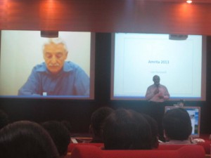
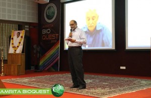
Dr. Hartwell is also adjunct faculty at Amrita University. He spoke to the delegates at Bioquest from his office in the US, over Amrita’s e-learning platform A-View. Given below are excerpts from his address.
I would like to address the young people in the audience. I know that many of you may have come to this meeting wondering, “How can I become a successful scientist? How can I prepare myself to make a contribution in this world?”
These questions are interesting to me also.
Believe it or not, I am still trying to be a successful scientist. That may surprise you since you probably think that a Nobel laureate must have found the answers. But the problem is that the answers to these questions change with time and the answers are different today than what they were when I began my career fifty years ago. The strategy of the 1960’s doesn’t work so well anymore. What is different now?
First, what we know now is much more. For example, by 1970, no genes from any organisms were sequenced. In 2013, we have the complete sequence of the human genome. Second, not only do we know much more today, accessing that knowledge is easy. Third, obtaining new information is much faster today.
Our rich understanding of science and technology is now needed to solve many serious problems. The human population has reached the size where we are utilizing all available resource of the planet. We are utilizing all of the agricultural land, all of the water, all of the forest and fishing resources. We are also polluting the planet that we live on.
We are polluting the land with fertilizers and pesticides; the oceans with acids and the atmosphere with carbon dioxide. We are using up top soil and ground water, thereby reducing our capacity to feed ourselves. We are using up petroleum, the energy source that our entire economy is dependent on. These are problems we were largely unaware of, fifty years ago. But these are problems that must be solved in your life times.
The big question facing your generation is, how can human beings live sustainably on planet earth. Your two broad goals on sustainability are 1) leave the planet as you first found it for your future generations; don’t use up the resources and don’t pollute the planet 2) everyone deserves to have an equal share of the earth’s resources.
Income strongly determines one’s opportunities in life. Many poor people succumb to chronic diseases and unhealthy environments. This inequality undermines our ability to live sustainably. We can’t ask the poor to leave the planet as they found it if they can’t support their families. Education, healthcare, employment are essential to having a sustainable society.
How can we be a successful scientist in 2013?
1. First choose a problem to solve
2. Ask questions to understand why it is not solved
3. Collaborate with those who can help
4. Develop a solution that works in the real worldChronic diseases are our major burden and this burden will get worse. Heart disease, diabetes, cancer, dementia and other diseases. The good news is that the chronic diseases are largely preventable and more easily curable if detected early. One question that attracts me is how can we detect disease earlier when it can be more easily cured?
Can we use our increasing knowledge in molecular biology to identify biomarkers for early disease detection?
We need to collaborate very closely with clinicians who care for patients to find out exactly where they need help.
I think if we apply our technology to important clinical questions we will actually save medical expenditure and be well on our way to making a great contribution to society.

H S M Kort, J-W J Lammers, S N W Vorrink, T Troosters
Introduction
Chronic Obstructive Pulmonary Disease (COPD) is a disabling airway disease with variable extrapulmonary effects that may contribute to disease severity in individual patients (Rabe et al. 2007). The world health organization predicts that COPD will become the third leading cause of death worldwide by 2030. Patients with COPD demonstrate reduced levels of spontaneous daily physical activity (DPA) compared with healthy controls (Pitta et al. 2005). This results in a higher risk of hospital admission and shorter survival (Pitta et al. 2006). Pulmonary rehabilitation can help to improve the DPA level, however, obtained benefits decline after 1–2 years (Foglio et al. 2007).
Purpose
In order to maintain DPA in COPD patients after rehabilitation, we developed a mobile phone application. This application measures DPA as steps per day, measured by the accelerometer of the smartphone, and shows the information to the patient via the display of the mobile phone. A physiotherapist can monitor the patient via a secure website where DPA measurements are visible for all patients. Here, DPA goals can be adjusted and text messages sent.
Method
Three pilot studies were performed with healthy students and COPD patients to test the application for usability, user friendliness and reliability with questionnaires and focus groups. Subjects also wore a validated accelerometer. For the Randomized Controlled Trial (RCT) 140 COPD patients will be recruited in Dutch physiotherapy practises. They will be randomised in an intervention group that receives the smartphone for 6 months and a control group. Measurements include lungfunction, dyspnea, and exercise capacity and are held at 0, 3, 6 and 12 months.
Results and Discussion
The application was found to be useful, easy to learn and use. Subjects had no problems with health care professionals seeing information on their physical activity performance. They do find it important to be able to determine who can see the information. Correlations between the accelerometer and the measurements on DPA of the smartphone for steps per hour were 0.69 and 0.70 for pilot studies 1 (students) and 2 (COPD patients) respectively. The version of the application in pilot study 3 contained an error, which made correlations with the accelerometer unusable. The RCT study is now being executed.
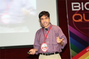
 Vural Özdemir, MD, Ph.D., DABCP
Co-Founder, DELSA Global, Seattle, WA, USA
Crowd-Funded Micro-Grants to Link Biotechnology and “Big Data” R&D to Life Sciences Innovation in India
Vural Özdemir, MD, PhD, DABCP1,2*
- Data-Enabled Life Sciences Alliance International (DELSA Global), Seattle, WA 98101, USA;
- Faculty of Management and Medicine, McGill University, Canada;
ABSTRACT
Aims: This presentation proposes two innovative funding solutions for linking biotechnology and “Big Data” R&D in India with artisan small scale discovery science, and ultimately, with knowledge-based innovation:
- crowd-funded micro-grants, and
- citizen philanthropy
These two concepts are new, and inter-related, and can be game changing to achieve the vision of biotechnology innovation in India, and help bridge local innovation with global science.
Background and Context: Biomedical science in the 21(st) century is embedded in, and draws from, a digital commons and “Big Data” created by high-throughput Omics technologies such as genomics. Classic Edisonian metaphors of science and scientists (i.e., “the lone genius” or other narrow definitions of expertise) are ill equipped to harness the vast promises of the 21(st) century digital commons. Moreover, in medicine and life sciences, experts often under-appreciate the important contributions made by citizen scholars and lead users of innovations to design innovative products and co-create new knowledge. We believe there are a large number of users waiting to be mobilized so as to engage with Big Data as citizen scientists-only if some funding were available. Yet many of these scholars may not meet the meta-criteria used to judge expertise, such as a track record in obtaining large research grants or a traditional academic curriculum vitae. This presentation will describe a novel idea and action framework: micro-grants, each worth $1000, for genomics and Big Data. Though a relatively small amount at first glance, this far exceeds the annual income of the “bottom one billion” – the 1.4 billion people living below the extreme poverty level defined by the World Bank ($1.25/day).
We will present two types of micro-grants. Type 1 micro-grants can be awarded through established funding agencies and philanthropies that create micro-granting programs to fund a broad and highly diverse array of small artisan labs and citizen scholars to connect genomics and Big Data with new models of discovery such as open user innovation. Type 2 micro-grants can be funded by existing or new science observatories and citizen think tanks through crowd-funding mechanisms described herein. Type 2 micro-grants would also facilitate global health diplomacy by co-creating crowd-funded micro-granting programs across nation-states in regions facing political and financial instability, while sharing similar disease burdens, therapeutics, and diagnostic needs. We report the creation of ten Type 2 micro-grants for citizen science and artisan labs to be administered by the nonprofit Data-Enabled Life Sciences Alliance International (DELSA Global, Seattle: http://www.delsaglobal.org). Our hope is that these micro-grants will spur novel forms of disruptive innovation and life sciences translation by artisan scientists and citizen scholars alike.
Address Correspondence to:
Vural Özdemir, MD, PhD, DABCP
Senior Scholar and Associate Professor
Faculty of Management and Medicine, McGill University
1001 Sherbrooke Street West
Montreal, Canada H3A 1G5
 Akhilesh Pandey, Ph.D.
Professor, Johns Hopkins University School of Medicine, Baltimore, USA
A draft map of the human proteome
We have generated a draft map of the human proteome through a systematic and comprehensive analysis of normal human adult tissues, fetal tissues and hematopoietic cells as an India-US initiative. This unique dataset was generated from 30 histologically normal adult tissues, fetal tissues and purified primary hematopoietic cells that were analyzed at high resolution in the MS mode and by HCD fragmentation in the MS/MS mode on LTQ-Orbitrap Velos/Elite mass spectrometers. This dataset was searched against a 6-frame translation of the human genome and RNA-Seq transcripts in addition to standard protein databases. In addition to confirming a large majority (>16,000) of the annotated protein-coding genes in humans, we obtained novel information at multiple levels: novel protein-coding genes, unannotated exons, novel splice sites, proof of translation of pseudogenes (i.e. genes incorrectly annotated as pseudogenes), fused genes, SNPs encoded in proteins and novel N-termini to name a few. Many proteins identified in this study were identified by proteomic methods for the first time (e.g. hypothetical proteins or proteins annotated based solely on their chromosomal location). We have generated a catalog of proteins that show a more tissue-restricted pattern of expression, which should serve as the basis for pursuing biomarkers for diseases pertaining to specific organs. This study also provides one of the largest sets of proteotypic peptides for use in developing MRM assays for human proteins. Identification of several novel protein-coding regions in the human genome underscores the importance of systematic characterization of the human proteome and accurate annotation of protein-coding genes. This comprehensive dataset will complement other global HUPO initiatives using antibody-based as well as MRM mass spectrometry-based strategies. Finally, we believe that this dataset will become a reference set for use as a spectral library as well as for interesting interrogations pertaining to biomedical as well as bioinformatics questions.

