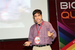
 Gaute Einevoll, Ph.D.
Professor of Physics, Department of Mathematical Sciences & Technology, Norwegian University of Life Sciences (UMB)
Multiscale modeling of cortical network activity: Key challenges
Gaute T. Einevoll Computational Neuroscience Group, Norwegian University of Life Sciences, 1432 Ås, Norway; Norwegian National Node of the International Neuroinformatics Coordinating Facility (INCF)
Several challenges must be met in the development of multiscale models of cortical network activity. In the presentation I will, based on ongoing work in our group (http://compneuro.umb.no/ ) on multiscale modeling of cortical columns, outline some of them. In particular,
-
Combined modeling schemes for neuronal, glial and vascular dynamics [1,2],
-
Development of sets of interconnected models describing system at different levels of biophysical detail [3-5],
-
Multimodal modeling, i.e., how to model what you can measure [6-12],
-
How to model when you don’t know all the parameters, and
-
Development of neuroinformatics tools and routines to do simulations efficiently and accurately [13,14].
References:
- L. Øyehaug, I. Østby, C. Lloyd, S.W. Omholt, and G.T. Einevoll: Dependence of spontaneous neuronal firing and depolarisation block on astroglial membrane transport mechanisms, J Comput Neurosci 32, 147-165 (2012)
- I. Østby, L. Øyehaug, G.T. Einevoll, E. Nagelhus, E. Plahte, T. Zeuthen, C. Lloyd, O.P. Ottersen, and S.W. Omholt: Astrocytic mechanisms explaining neural-activity-induced shrinkage of extraneuronal space, PLoS Comp Biol 5, e1000272 (2009)
- T. Heiberg, B. Kriener, T. Tetzlaff, A. Casti, G.T. Einevoll, and H.E. Plesser: Firing-rate models can describe the dynamics of the retina-LGN connection, J Comput Neurosci (2013)
- T. Tetzlaff, M. Helias, G.T. Einevoll, and M. Diesmann: Decorrelation of neural-network activity by inhibitory feedback, PLoS Comp Biol 8, e10002596 (2012).
- E. Nordlie, T. Tetzlaff, and G.T. Einevoll: Rate dynamics of leaky integrate-and-fire neurons with strong synapses, Frontiers in Comput Neurosci 4, 149 (2010)
- G.T. Einevoll, F. Franke, E. Hagen, C. Pouzat, K.D. Harris: Towards reliable spike-train recording from thousands of neurons with multielectrodes, Current Opinion in Neurobiology 22, 11-17 (2012)
- H. Linden, T Tetzlaff, TC Potjans, KH Pettersen, S Grun, M Diesmann, GT Einevoll: Modeling the spatial reach of LFP, Neuron 72, 859-872 (2011).
- H. Linden, K.H. Pettersen, and G.T. Einevoll: Intrinsic dendritic filtering gives low-pass power spectra of local field potentials, J Computational Neurosci 29, 423-444 (2010)
- K.H. Pettersen and G.T. Einevoll: Amplitude variability and extracellular low-pass filtering of neuronal spikes, Biophysical Journal 94, 784-802 (2008).
- K.H. Pettersen, E. Hagen, and G.T. Einevoll: Estimation of population firing rates and current source densities from laminar electrode recordings, J Comput Neurosci 24, 291-313 (2008).
- K. Pettersen, A. Devor, I. Ulbert, A.M. Dale and G.T. Einevoll. Current-source density estimation based on inversion of electrostatic forward solution: Effects of finite extent of neuronal activity and conductivity discontinuities, Journal of Neuroscience Methods 154, 116-133 (2006).
- G.T. Einevoll, K. Pettersen, A. Devor, I. Ulbert, E. Halgren and A.M. Dale: Laminar Population Analysis: Estimating firing rates and evoked synaptic activity from multielectrode recordings in rat barrel cortex, Journal of Neurophysiology 97, 2174-2190 (2007).
- LFPy: A tool for simulation of extracellular potentials (http://compneuro.umb.no)
- E. Nordlie, M.-O. Gewaltig, H. E. Plesser: Towards reproducible descriptions of neuronal network models, PLoS Comp Biol 5, e1000456 (2009).
 Rohit Manchanda, Ph.D.
Professor, Biomedical Engineering Group, IIT-Bombay, India
Modelling the syncytial organization and neural control of smooth muscle: insights into autonomic physiology and pharmacology
We have been studying computationally the syncytial organization and neural control of smooth muscle in order to help explain certain puzzling findings thrown up by experimental work. This relates in particular to electrical signals generated in smooth muscles, such as synaptic potentials and spikes, and how these are explicable only if three-dimensional syncytial biophysics are taken fully into account. In this talk, I shall provide an illustration of outcomes and insights gleaned from such an approach. I shall first describe our work on the mammalian vas deferens, in which an analysis of the effects of syncytial coupling led us to conclude that the experimental effects of a presumptive gap junction uncoupler, heptanol, on synaptic potentials were incompatible with gap junctional block and could best be explained by a heptanol-induced inhibition of neurotransmitter release, thus compelling a reinterpretation of the mechanism of action of this agent. I shall outline the various lines of evidence, based on indices of syncytial function, that we adduced in order to reach this conclusion. We have now moved on to our current focus on urinary bladder biophysics, where the questions we aim to address are to do with mechanisms of spike generation. Smooth muscle cells in the bladder exhibit spontaneous spiking and spikes occur in a variety of distinct shapes, making their generation problematic to explain. We believe that the variety in shapes may owe less to intrinsic differences in spike mechanism (i.e., in the complement of ion channels participating in spike production) and more to features imposed by syncytial biophysics. We focus especially on the modulation of spike shape in a 3-D coupled network by such factors as innervation pattern, propagation in a syncytium, electrically finite bundles within and between which the spikes spread, and some degree of pacemaker activity by a sub-population of the cells. I shall report two streams of work that we have done, and the tentative conclusions these have enabled us to reach: (a) using the NEURON environment, to construct the smooth muscle syncytium and endow it with synaptic drive, and (b) using signal-processing approaches, towards sorting and classifying the experimentally recorded spikes.
 Leland H. Hartwell Ph.D.
2001 Nobel Laureate, Physiology & Medicine
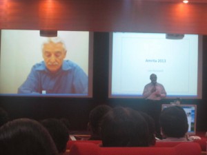
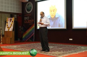
Dr. Lee Hartwell received the 2001 Nobel Prize in Physiology / Medicine for his discovery of protein molecules that control the division of cells. He was the President and Director of the Fred Hutchinson Cancer Research Center in Seattle, Washington before moving to Arizona State University’s Center for Sustainable Health.
Dr. Hartwell is also adjunct faculty at Amrita University. He spoke to the delegates at Bioquest from his office in the US, over Amrita’s e-learning platform A-View. Given below are excerpts from his address.
I would like to address the young people in the audience. I know that many of you may have come to this meeting wondering, “How can I become a successful scientist? How can I prepare myself to make a contribution in this world?”
These questions are interesting to me also.
Believe it or not, I am still trying to be a successful scientist. That may surprise you since you probably think that a Nobel laureate must have found the answers. But the problem is that the answers to these questions change with time and the answers are different today than what they were when I began my career fifty years ago. The strategy of the 1960’s doesn’t work so well anymore. What is different now?
First, what we know now is much more. For example, by 1970, no genes from any organisms were sequenced. In 2013, we have the complete sequence of the human genome. Second, not only do we know much more today, accessing that knowledge is easy. Third, obtaining new information is much faster today.
Our rich understanding of science and technology is now needed to solve many serious problems. The human population has reached the size where we are utilizing all available resource of the planet. We are utilizing all of the agricultural land, all of the water, all of the forest and fishing resources. We are also polluting the planet that we live on.
We are polluting the land with fertilizers and pesticides; the oceans with acids and the atmosphere with carbon dioxide. We are using up top soil and ground water, thereby reducing our capacity to feed ourselves. We are using up petroleum, the energy source that our entire economy is dependent on. These are problems we were largely unaware of, fifty years ago. But these are problems that must be solved in your life times.
The big question facing your generation is, how can human beings live sustainably on planet earth. Your two broad goals on sustainability are 1) leave the planet as you first found it for your future generations; don’t use up the resources and don’t pollute the planet 2) everyone deserves to have an equal share of the earth’s resources.
Income strongly determines one’s opportunities in life. Many poor people succumb to chronic diseases and unhealthy environments. This inequality undermines our ability to live sustainably. We can’t ask the poor to leave the planet as they found it if they can’t support their families. Education, healthcare, employment are essential to having a sustainable society.
How can we be a successful scientist in 2013?
1. First choose a problem to solve
2. Ask questions to understand why it is not solved
3. Collaborate with those who can help
4. Develop a solution that works in the real worldChronic diseases are our major burden and this burden will get worse. Heart disease, diabetes, cancer, dementia and other diseases. The good news is that the chronic diseases are largely preventable and more easily curable if detected early. One question that attracts me is how can we detect disease earlier when it can be more easily cured?
Can we use our increasing knowledge in molecular biology to identify biomarkers for early disease detection?
We need to collaborate very closely with clinicians who care for patients to find out exactly where they need help.
I think if we apply our technology to important clinical questions we will actually save medical expenditure and be well on our way to making a great contribution to society.
 Nader Pourmand, Ph.D.
Director, UCSC Genome Technology Center,University of California, Santa Cruz
Biosensor and Single Cell Manipulation using Nanopipettes
Approaching sub-cellular biological problems from an engineering perspective begs for the incorporation of electronic readouts. With their high sensitivity and low invasiveness, nanotechnology-based tools hold great promise for biochemical sensing and single-cell manipulation. During my talk I will discuss the incorporation of electrical measurements into nanopipette technology and present results showing the rapid and reversible response of these subcellular sensors to different analytes such as antigens, ions and carbohydrates. In addition, I will present the development of a single-cell manipulation platform that uses a nanopipette in a scanning ion-conductive microscopy technique. We use this newly developed technology to position the nanopipette with nanoscale precision, and to inject and/or aspirate a minute amount of material to and from individual cells or organelle without comprising cell viability. Furthermore, if time permits, I will show our strategy for a new, single-cell DNA/ RNA sequencing technology that will potentially use nanopipette technology to analyze the minute amount of aspirated cellular material.
 Satheesh Babu T. G., Ph.D.
Associate Professor, Department of Sciences, School of Engineering, Amrita University, Coimbatore, India
Nanomaterials for ‘enzyme-free’ biosensing
Enzyme based sensors have many draw backs such as poor storage stability, easily affected by the change in pH and temperature and involves complicated enzyme immobilization procedures. To address this limitation, an alternative approach without the use of enzyme, “non-enzymatic” has been tried recently. Choosing the right catalyst for direct electrochemical oxidation / reduction of a target molecule is the key step in the fabrication of non-enzymatic sensors.
Non-enzymatic sensors for glucose, creatinine, vitamins and cholesterol are fabricated using different nanomaterials, such as nanotubes, nanowires and nanoparticles of copper oxide, titanium dioxide, tantalum oxide, platinum, gold and graphenes. These sensors selectively catalyse the targeted analyte with very high sensitivity. These nanomaterials based sensors combat the drawbacks of enzymatic sensors.

Anupama Natarajan, James Hickman and Peter Molnar
Novel Cell-Based Biosensors for High Throughput Toxin Detection and Drug Screening Applications
Over the last decade there has been focus on the development of cellbased biosensors to detect environmental toxins or to combat the threats of biological warfare. These sensors have been shown to have multiple applications including understanding function and behaviour at the cellular and tissue levels, in cell electrophysiology and as drug screening tools that can eliminate animal testing. These factors make the development of cell-based biosensors into high throughput systems a priority in pharmacological, environmental and defence industries (Pancrazio J J et al. 1999, Kang G et al. 2009, Krinke D et al. 2009). We have developed a high through-put in vitro cell-silicon hybrid platform that could be used to analyze both cell function and response to various toxins and drugs. Our hypothesis was that by utilizing surface modification to provide external guidance cues as well as optimal growth conditions for different cell types (Cardiac and Neuronal), we could enhance the information output and content of such a system. An intrinsic part of this study was to create ordered or patterned functional networks of cells on Micro-electrode arrays (MEA). Such engineered networks had a two-fold purpose in that they not only aided in a more accurate analysis of cell response and cell and tissue behaviour, but also increased the efficiency of the system by increasing the connectivity and placement of the cells over the recording electrodes. Here we show the response of this system to various toxins and drugs and the measurement of several vital cardiac parameters like conduction velocity and refractory period (Natarajan A et al. 2011)
 Akhilesh Pandey, Ph.D.
Professor, Johns Hopkins University School of Medicine, Baltimore, USA
A draft map of the human proteome
We have generated a draft map of the human proteome through a systematic and comprehensive analysis of normal human adult tissues, fetal tissues and hematopoietic cells as an India-US initiative. This unique dataset was generated from 30 histologically normal adult tissues, fetal tissues and purified primary hematopoietic cells that were analyzed at high resolution in the MS mode and by HCD fragmentation in the MS/MS mode on LTQ-Orbitrap Velos/Elite mass spectrometers. This dataset was searched against a 6-frame translation of the human genome and RNA-Seq transcripts in addition to standard protein databases. In addition to confirming a large majority (>16,000) of the annotated protein-coding genes in humans, we obtained novel information at multiple levels: novel protein-coding genes, unannotated exons, novel splice sites, proof of translation of pseudogenes (i.e. genes incorrectly annotated as pseudogenes), fused genes, SNPs encoded in proteins and novel N-termini to name a few. Many proteins identified in this study were identified by proteomic methods for the first time (e.g. hypothetical proteins or proteins annotated based solely on their chromosomal location). We have generated a catalog of proteins that show a more tissue-restricted pattern of expression, which should serve as the basis for pursuing biomarkers for diseases pertaining to specific organs. This study also provides one of the largest sets of proteotypic peptides for use in developing MRM assays for human proteins. Identification of several novel protein-coding regions in the human genome underscores the importance of systematic characterization of the human proteome and accurate annotation of protein-coding genes. This comprehensive dataset will complement other global HUPO initiatives using antibody-based as well as MRM mass spectrometry-based strategies. Finally, we believe that this dataset will become a reference set for use as a spectral library as well as for interesting interrogations pertaining to biomedical as well as bioinformatics questions.


