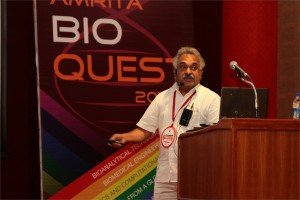
 K. P. Mohanakumar, Ph.D.
Chief Scientist, Cell Biology & Physiology Division, Indian Institute of Chemical Biology, Kolkata
Neuroprotective and neurodestructive effects of Ayurvedic drug constituents: Parkinson’s disease
The present study reports the good and the bad entities in an Indian traditional medicine used for treating Parkinson’s disease (PD). A prospective clinical trial on the effectiveness of Ayurvedic medication in a population of PD patients revealed significant benefits, which has been attributed to L-DOPA present in the herbs [1]. Later studies revealed better benefits with one of the herbs alone, compared to pure L-DOPA in a clinical trial conducted in UK [2], and in several studies conducted on animal models of PD in independent laboratories world over [3-5]. We have adapted strategies to segregate molecules from the herb, and then carefully removed L-DOPA contained therein, and tested each of these sub-fractions for anti-PD activity in 1-methyl-4-phenyl-1,2,3,6-tetrahydropyridine, rotenone and 6-hydroxydopamine -induced parkinsonian animal models, and transgenic mitochondrial cybrids. We report here two classes of molecules contained in the herb, one of which possessed severe pro-parkinsonian (phenolic amine derivatives) and the other having excellent anti-parkinsonian potential (substituted tetrahydroisoquinoline derivatives). The former has been shown to cause severe dopamine depletion in the striatum of rodents, when administered acutely or chronically. It also caused significant behavioral aberrations, leading to anxiety and depression [6]. The latter class of molecules administered in PD animal model [7], caused reversal of behavioral dysfunctions and significant attenuation of striatal dopamine loss. These effects were comparable or better than the effects of the anti-PD drugs, selegiline or L-DOPA. The mechanism of action of the molecule has been found to be novel, at the postsynaptic receptor signaling level, as well as cellular α-synuclein oligomerization and specifically at mitochondria. The molecule helped in maintaining mitochondrial ETC complex activity and stabilized cellular respiration, and mitochondrial fusion-fission machinery with specific effect on the dynamin related protein 1. Although there existed significant medical benefits that could be derived to patients due to the synergistic actions of several molecules present in a traditional preparation, accumulated data in our hands suggest complicated mechanisms of actions of Ayurvedic medication. Our results also provide great hope for extracting, synthesizing and optimizing the activity of anti-parkinsonian molecules present in traditional Ayurvedic herbs, and for designing novel drugs with novel mechanisms of action.
- N, Nagashayana, P Sankarankutty, MRV Nampoothiri, PK Mohan and KP Mohanakumar, J Neurol Sci. 176, 124-7, 2000.
- Katzenschlager R, Evans A, Manson A, Patsalos PN, Ratnaraj N, Watt H, Timmermann L, Van der Giessen R, Lees AJ. J Neurol Neurosurg Psychiatry.75, 1672-7, 2004.
- Manyam BV, Dhanasekaran M, Hare TA. Phytother Res. 18, 706-12, 2004.
- Kasture S, Pontis S, Pinna A, Schintu N, Spina L, Longoni R, Simola N, Ballero M, Morelli M. Neurotox Res. 15, 111-22, 2009.
- Lieu CA, Kunselman AR, Manyam BV, Venkiteswaran K, Subramanian T. Parkinsonism Relat Disord.16, 458-65, 2010.
- T Sengupta and KP Mohanakumar, Neurochem Int. 57, 637-46, 2010.
- T Sengupta, J Vinayagam, N Nagashayana, B Gowda, P Jaisankar and KP Mohanakumar, Neurochem Res 36, 177-86, 2011

Binu K Aa, Jem Prabhakarb, Thara Sc and Lakshmi Sd,∗
aDepartment of Clinical Diagnostics Services and Translational Research, Malabar Cancer Centre, Thalassery, Kerala, India.
bDivision of Surgical Oncology, Division of Pathology
dDivision of Cancer Research, Regional Cancer Centre, Thiruvananthapuram, Kerala, India.
Introduction
AIB1, a member of the nuclear co activators, promotes the transcriptional activity of multiple nuclear receptors such as the ER and other transcription factors. Chemokines produced by stromal cells have potential to influence ERα-positive breast cancer progression to metastasis. CXCR4 is the physiological receptor for SDF1, together shown to stimulate the chemotactic and invasive behavior of breast cancer cells to serve as a homing mechanism to sites of metastasis. We propose that over expression of AIB1 in breast cancer cells leads to increased SDF1 and CXCR4 expression, which induces invasion and metastasis of cancer cells.
Materials and Methods
Breast tumor and normal breast tissues from patients in Regional Cancer Centre, Thiruvananthapuram were used for study. The modulatory effect of AIB1 was studied in MCF-7 cells with AIB1 siRNA transfection along with treatment of 17β-Estradiol (E2), 4-hydroxytamoxifen (4OHT), combinations of E2 and 4OHT. The gene expression pattern and protein localization were assessed by RT-PCR and immunofluorescence microscopy respectively. The metastatic and invasive properties were assessed by wound healing assay. Quantitative colocalization analyses were done to assess the association of proteins using Pearson’s correlation coefficient.
Result and Conclusion
The mRNA and protein level expression of AIB1, CXCR4 and SDF1 were higher in tumor samples than in normal samples. AIB1 was localized to the nuclei whereas CXCR4 and SDF1 immunoreactivity were observed in the cytoplasm and to a lesser extent in the nuclei of tumor epithelial cells. In tumor samples the gene level expressions of AIB1 showed significant positive correlations with SDF1(r = 0.213, p = 0.018). CXCR4 showed significant positive correlation with SDF1 in gene (r = 0.498, p = 0.000) and protein levels(r = 0.375, p = 0.002). Quantitative colocalization analyses showed a marked reduction in expression of CXCR4 and SDF1 in siAIB1MCF-7 cells than MCF-7 cells with different treatment groups. Wound healing assay shows reduced wound healing in siAIB1 treated MCF-7 cells.
In recent years, targeting specific cancer pathways and key molecules to arrest tumor growth and achieve tumor eradication have proven a challenge; due to acquired resistance and homing of cancer cells to various metastatic sites. The present study revealed that silencing AIB1 can prevent the over expression of SDF1 and CXCR4. Co activator levels determine the basal and estrogen-inducible expression of SDF1, a secreted protein that controls breast cancer cell proliferation and invasion through autocrine and paracrine mechanisms (Hall et al. 2003). The effects of CXCR4 overexpression has been correlated with SDF1 mediated activation of downstream signaling via ERK1/2 and p38 MAPK and with an enhancement of ER-mediated gene expression (Rhodes et al. 2011). It is possible that over expression of AIB1 as a stimulant involved in the expression of CXCR4 might up-regulate the expression of prometastatic and angiogenic genes. Thus based on these observations it can be concluded that SDF1/CXCR4 overexpression, with significant association with AIB1 expression, itself contribute to the development of mammary cancer and metastatic progression.


