 Ashok Pandey, Ph.D.
Scientist F & Head, Biotechnology Division, National Institute for Interdisciplinary Science and Technology-CSIR), Thiruvananthapuram, India
Alternative renewable resources: Issues and perspectives for India – the case of transport fuels
With the increase in the urbanization way of life and also more and more dependence on materialistic life, there is substantial growing demand for the energy. The science and technological policy of the India has looked several avenues to fulfill this demand through alternative resources such as solar energy, wind energy, tidal energy, bioenergy, etc. The demand for the transport sector is largely met through the import (~70%). Biofuels, in particular bioethanol from lignocellulosic biomass offer attractive possibilities in this regard.
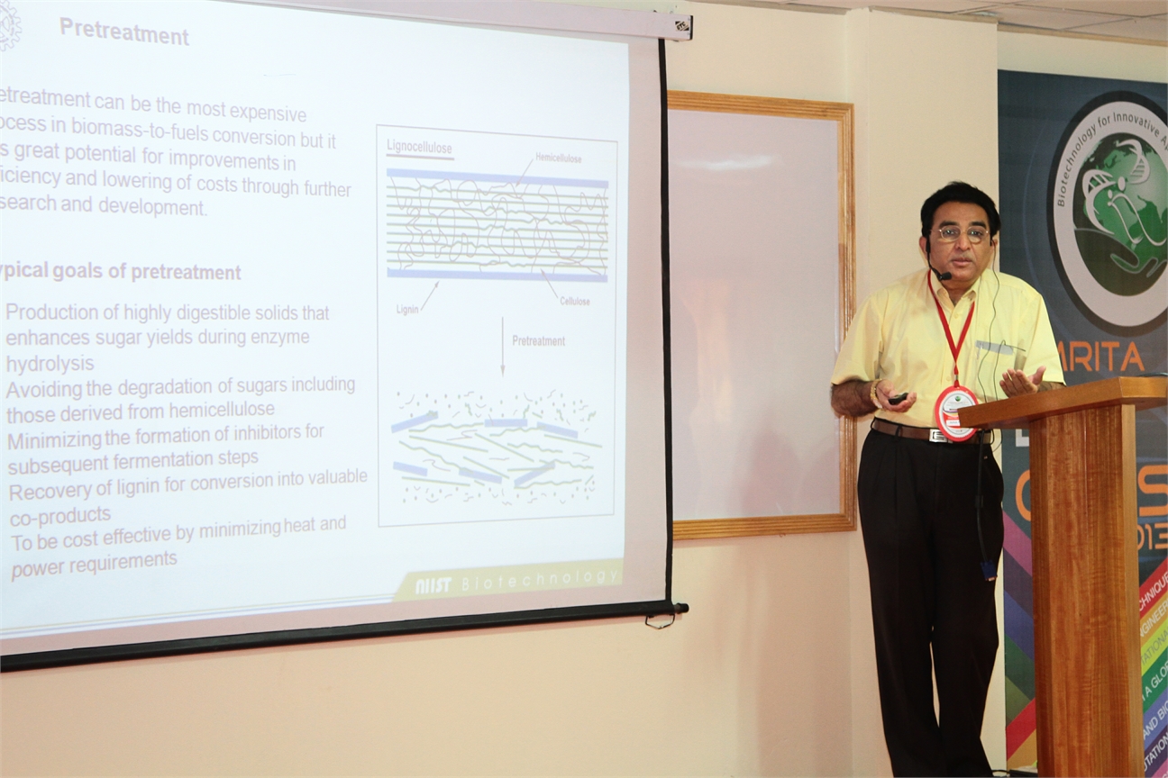
The sugar platform which generates ethanol is considered to be the most valuable solution to the transport fuel demand. Bioethanol can be generated from grains as well as from lignocellulosic plant material by their saccharification to sugars and subsequent fermentation of the sugars to produce ethanol. Bio-ethanol as a transportation fuel is attractive since it is more energy efficient than gasoline and produces less emissions. The benefits of developing biomass to ethanol technology(s) include: increased national energy security, reduction in GHG emissions, use of renewable resources, economic benefits and creation of employment and the foundation of a carbohydrate based chemical industry. However, the utilization of lignocellulosic biomass for fuel generation has not been given the sort of attention it ought to receive. It is known that the technology for ethanol production from biomass has to evolve greatly for an economical commercial scale utilization of the renewable biomass resources. Biomass requires extensive processing involving multiple steps for hydrolysis and fermentation of the raw material for producing ethanol. Feed stock availability, pretreatment, saccharification, fermentation and ethanol recovery are all factors which influence the production of ethanol and which needs R&D efforts for overall improvement of the production economics.
Bioconversion of lignocellulosic biomass (LB) can contribute significantly to the production of organic chemicals also. LB is also considered to be the only foreseeable source of energy. LB is mainly composed of (dry wt basis): cellulose, 40-60; hemicellulose, 20-40; and lignin, 10-25%. Most efficient method of biomass hydrolysis is through enzymatic saccharification5 using cellulases and hemicellulases. Fungal cellulases (FCs) have proved to be a better candidate than other microbial cellulases, with their secreted free cellulase complexes comprising all three components of cellulase [endoglucanases, exoglucanases and cellobiases (glucosidases).
The Centre for Biofuels at NIIST, Trivandrum, India aims ultimately to develop technologies and processes which will address the nation’s need for making fuel ethanol from the renewable resource: biomass. It is proposed to direct R&D activities at the major requirements of a biomass-ethanol technology, which include production of cellulases, hydrolysis of biomass, and ethanol fermentation. Viable technologies for each of these processes will contribute to the overall process development for fuel alcohol production from cheap and renewable biomass resources.
The lecture would present perspectives on bioethanol from lignocellulosic feedstocks.
References
- Biofuels- Alternative Feedstocks and Conversion Processes, Editors- Ashok Pandey, C Larroche, SC Ricke, CG Dussap & E Gnansounou, Academic Press, Elsevier Inc; San Diego, USA, p629 (2011) ISBN: 978-0-12-385099-7
- Handbook of Plant-Based Biofuels, Editor- Ashok Pandey, CRC Press, Francis & Taylors, Boca Raton, USA, p 297 (2008) ISBN 978-q-5602-2175-3
- Biofuels II, Special issue of Journal of Scientific & Industrial Research, Guest Editors- E Gnansounou, C Larroche and Ashok Pandey, 67(11), 837-1040 (2008) ISSN: 0022-4456
- Biofuels, Special issue of Journal of Scientific & Industrial Research, Guest Editors- C Larroche and Ashok Pandey, 64(11), 797-988 (2005) ISSN: 0022-4456
 Claudia AM Wheeler-Kingshott, Ph.D.
University Reader in Magnetic Resonance Physics, Department of Neuroinflammation, UCL Institute of Neurology, London, UK
Abstract
Detecting neuronal activity in vivo non-invasively is possible with a number of techniques. Amongst these, in 1990 functional magnetic resonance imaging (fMRI) was proposed as a technique that has a great ability to spatially map brain activity by exploiting the blood oxygenation level dependent (BOLD) contrast mechanism [1, 2]. In fact, neuronal activation triggers a demand for oxygen and induces a localised increase in blood flow and blood volume, which actually exceeds the metabolic needs. This in turns causes an increase of oxyhaemoglobin in the venous compartment, which is a transient phenomenon and is accompanied by a transient change (decrease) in the concentration of deoxyhaemoglobin. Due to its paramagnetic properties, the amount of deoxyhaemoglobin present in the venous blood affects the local magnetic field seen by the spins (protons) and determines the local properties of the MR signal. A decrease in deoxyhaemoglobin during neuronal activity, therefore, induces local variations of this magnetic field that increases the average transverse relaxation time of tissue, measured via the T2* parameter [3]. This means that there is an increase of the MR signal (of the order of a few %, typically <5%) linked to metabolic changes happening during brain function. Activation can be inferred at different brain locations by performing tasks while acquiring the MR signal and comparing periods of rest to periods of activity.
The macroscopic changes of the BOLD signal are well characterised, while the reason for the increased blood supply, exceeding demands, needs further thoughts. Here we will discuss two approaches for explaining the BOLD phenomenon, one that links it to adenosine triphosphate production [4] and enzyme saturation, the other that relates it to the very slow diffusion of oxygen through the blood-brain-barrier with a consequent compensatory high demand of oxygen [5]. Some evidence of restricted oxygen diffusion has been shown by means of hypercapnia [6], although it is not excluded that both mechanisms may be present.
Overall, the BOLD signal changes theory and its physiological basis will be presented and discussed.
References
- Ogawa, S., et al., Brain magnetic resonance imaging with contrast dependent on blood oxygenation. Proc Natl Acad Sci U S A, 1990. 87(24): p. 9868-72.
- Kwong, K.K., et al., Dynamic magnetic resonance imaging of human brain activity during primary sensory stimulation. Proc Natl Acad Sci U S A, 1992. 89(12): p. 5675-9.
- Bandettini PA, et al. Spin-echo and gradient-echo EPI of human brain activation using BOLD contrast: a comparative study at 1.5 T. NMR Biomed. 1994 Mar;7(1-2):12-20
- Fox, P.T., et al., Nonoxidative glucose consumption during focal physiologic neural activity. Science, 1988. 241(4864): p. 462-4.
- Gjedde, A., et al. Reduction of functional capillary density in human brain after stroke. J Cereb Blood Flow Metab, 1990. 10(3): p. 317-26.
- Hoge, R.D., et al., Linear coupling between cerebral blood flow and oxygen consumption in activated human cortex. Proc Natl Acad Sci U S A, 1999. 96(16): p. 9403-8.
 Gokul Das, Ph.D.
Co-Director, Breast Disease Site Research Group, Roswell Park Cancer Institute, Buffalo, NY
Probing Estrogen Receptor−Tumor Suppressor p53 Interaction in Cancer: From Basic Research to Clinical Trial
Tumor suppressor p53 and estrogen receptor have opposite roles in the onset and progression of breast cancer. p53 responds to a variety of cellular of stresses by restricting the proliferation and survival of abnormal cells. Estrogen receptor plays an important role in normal mammary gland development and the preservation of adult mammary gland function; however, when deregulated it becomes abnormally pro-proliferative and greatly contributes to breast tumorigenesis. The biological actions of estrogens are mediated by two genetically distinct estrogen receptors (ERs): ER alpha and ER beta. In addition to its expression in several ER alpha-positive breast cancers and normal mammary cells, ER beta is usually present in ER alpha-negative cancers including triple-negative breast cancer. In spite of genetically being wild type, why p53 is functionally debilitated in breast cancer has remained unclear. Our recent finding that ER alpha binds directly to p53 and inhibits its function has provided a novel mechanism for inactivating genetically wild type p53 in human cancer. Using a combination of proliferation and apoptosis assays, RNAi technology, quantitative chromatin immunoprecipitation (qChIP), and quantitative real-time PCR (qRT-PCR), in situ proximity ligation assay (PLA), and protein expression analysis in patient tissue micro array (TMA), we have demonstrated binding of ER alpha to p53 and have delineated the domains on both the proteins necessary for the interaction. Importantly, ionizing radiation inhibits the ER-p53 interaction in vivo both in human cancer cells and human breast tumor xenografts in mice. In addition, antiestrogenstamoxifen and faslodex/fulvestrant (ICI 182780) disrupt the ER-p53 interaction and counteract the repressive effect of ER alpha on p53, whereas 17β-estradiol (E2) enhances the interaction. Intriguingly, E2 has diametrically opposite effects on corepressor recruitment to a p53-target gene promoter versus a prototypic ERE-containing promoter. Thus, we have uncovered a novel mechanism by which estrogen could be providing a strong proliferative advantage to cells by dual mechanisms: enhancing expression of ERE-containing pro-proliferative genes while at the same time inhibiting transcription of p53-dependent anti-proliferative genes. Consistently, ER alpha enhances cell cycle progression and inhibits apoptosis of breast cancer cells. Correlating with these observations, our retrospective clinical study shows that presence of wild type p53 in ER-positive breast tumors is associated with better response to tamoxifen therapy. These data suggest ER alpha-p53 interaction could be one of the mechanisms underlying resistance to tamoxifen therapy, a major clinical challenge encountered in breast cancer patients. We have launched a prospective clinical trial to analyze ER-p53 interaction in breast cancer patient tumors at Roswell Park Cancer Institute. Our more recent finding that ER beta has opposite functions depending on the mutational status of p53 in breast cancer cells is significant in understanding the hard-to-treat triple-negative breast cancer and in developing novel therapeutic strategies against it. Our integrated approach to analyze ER-p53 interaction at the basic, translational, and clinical research levels has major implications in the diagnosis, prognosis, and treatment of breast cancer.