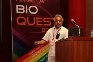
 Hideaki Nagase, Ph.D.
Kennedy Institute of Rheumatology-Centre for Degenerative Diseases, University of Oxford, UK
Osteoarthritis: diagnosis, treatment and challenges
Hideaki Nagase1, Ngee Han Lim1, George Bou-Gharios1, Ernst Meinjohanns2 and Morten Meldal3
- Kennedy Institute of Rheumatology, Nuffield Department of Orthopaedics, Rheumatology and Musculoskeletal Sciences, University of Oxford, London, W6 8LH UK
- Carlsberg Laboratory, Copenhagen, Denmark,
- Nano-Science Center, Department of Chemistry, University of Copenhagen, Denmark
Osteoarthritis (OA) is the most prevalent age-related degenerative joint disease. With the expanding ageing population, it imposes a major socio-economic burden on society. A key feature of OA is a gradual loss of articular cartilage and deformation of bone, resulting in the impairment of joint function. Currently, there is no effective disease-modifying treatment except joint replacement surgery. There are many possible causes of cartilage loss (e.g. mechanical load, injury, reactive oxygen species, aging, etc.) and etiological factors (obesity, genetics), but the degradation of cartilage is primarily caused by elevated levels of active metalloproteinases. It is therefore attractive to consider proteinase inhibitors as potential therapeutics. However, there are several hurdles to overcome, namely early diagnosis and continuous monitoring of the efficacy of inhibitor therapeutics. We are therefore aiming at developing non-invasive probes to detect cartilage degrading metalloproteinase activities.
We have designed in vivo imaging probes to detect MMP-13 (collagenase 3) activity that participates in OA by degrade cartilage collagen II and MMP-12 (macrophage elastase) activity involved in inflammatory arthritis. These activity-based probes consist of a peptide that is selectively cleaved by the target proteinase, a near-infrared fluorophore and a quencher. The probe’s signal multiplies upon proteolysis. They were first used to follow the respective enzyme activity in vivo in the mouse model of collagen-induced arthritis and we found MMP-12 activity probe (MMP12AP) activation peaked at 5 days after onset of the disease, whereas MMP13AP activation was observed at 10-15 days. The in vivo activation of these probes was inhibited by specific low molecule inhibitors. We proceeded to test both probes in the mouse model of OA induced by the surgical destabilization of medial meniscus of the knee joints. In this model, degradation of knee cartilage is first detected histologically 6 weeks after surgery with significant erosion detectable at 8 weeks. Little activation of MMP12AP was detected, which was expected, as macrophage migration is not obvious in OA. MMP13AP, on the other hand, was significantly activated in the operated knee at 6 weeks compared with the non-operated contralateral knee, but there were no significant differences between the operated and sham-operated knees. At 8 weeks, however, the signals in the operated knees were significantly higher than both the contralateral and sham-operated controls. Activation of aggrecanases and MMP-13 are observed before structural changes of cartilage. We are therefore currently improving the MMP-13 probe for earlier detection by attaching it to polymers that are retained in cartilage.
 Egidio D’Angelo, MD, Ph.D.
Full Professor of Physiology & Director, Brain Connectivity Center, University of Pavia, Italy
Realistic modeling: new insight into the functions of the cerebellar network
Realistic modeling is an approach based on the careful reconstruction of neurons synapses starting from biological details at the molecular and cellular level. This technique, combined with the connection topologies derived from histological measurements, allows the reconstruction of precise neuronal networks. Finally, the advent of specific software platforms (PYTHON-NEURON) and of super-computers allows large-scale network simulation to be performed in reasonable time. This approach inverts the logics of older theoretical models, which anticipated an intuition on how the network might work. In realistic modeling, network properties “emerge” from the numerous biological properties embedded into the model.
This approach is illustrated here through an outstanding application of realistic modeling to the cerebellar cortex network. The neurons (over 105) are reproduced at a high level of detail generating non-linear network effects like population oscillations and resonance, phase-reset, bursting, rebounds, short-term and long-term plasticity, spatiotemporal redistrbution of input patterns. The model is currently being used in the context of he HUMAN BRAIN PROJECT to investigate the cerebellar network function.
Correspondence should be addressed to
Dr. EgidioD’Angelo,
Laboratory of Neurophysiology
Via Forlanini 6, 27100 Pavia, Italy
Phone: 0039 (0) 382 987606
Fax: 0039 (0) 382 987527
dangelo@unipv.it
Acknowledgments
This work was supported by grants from European Union to ED (CEREBNET FP7-ITN238686, REALNET FP7-ICT270434) and by grants from the Italian Ministry of Health to ED (RF-2009-1475845).
 K. P. Mohanakumar, Ph.D.
Chief Scientist, Cell Biology & Physiology Division, Indian Institute of Chemical Biology, Kolkata
Neuroprotective and neurodestructive effects of Ayurvedic drug constituents: Parkinson’s disease
The present study reports the good and the bad entities in an Indian traditional medicine used for treating Parkinson’s disease (PD). A prospective clinical trial on the effectiveness of Ayurvedic medication in a population of PD patients revealed significant benefits, which has been attributed to L-DOPA present in the herbs [1]. Later studies revealed better benefits with one of the herbs alone, compared to pure L-DOPA in a clinical trial conducted in UK [2], and in several studies conducted on animal models of PD in independent laboratories world over [3-5]. We have adapted strategies to segregate molecules from the herb, and then carefully removed L-DOPA contained therein, and tested each of these sub-fractions for anti-PD activity in 1-methyl-4-phenyl-1,2,3,6-tetrahydropyridine, rotenone and 6-hydroxydopamine -induced parkinsonian animal models, and transgenic mitochondrial cybrids. We report here two classes of molecules contained in the herb, one of which possessed severe pro-parkinsonian (phenolic amine derivatives) and the other having excellent anti-parkinsonian potential (substituted tetrahydroisoquinoline derivatives). The former has been shown to cause severe dopamine depletion in the striatum of rodents, when administered acutely or chronically. It also caused significant behavioral aberrations, leading to anxiety and depression [6]. The latter class of molecules administered in PD animal model [7], caused reversal of behavioral dysfunctions and significant attenuation of striatal dopamine loss. These effects were comparable or better than the effects of the anti-PD drugs, selegiline or L-DOPA. The mechanism of action of the molecule has been found to be novel, at the postsynaptic receptor signaling level, as well as cellular α-synuclein oligomerization and specifically at mitochondria. The molecule helped in maintaining mitochondrial ETC complex activity and stabilized cellular respiration, and mitochondrial fusion-fission machinery with specific effect on the dynamin related protein 1. Although there existed significant medical benefits that could be derived to patients due to the synergistic actions of several molecules present in a traditional preparation, accumulated data in our hands suggest complicated mechanisms of actions of Ayurvedic medication. Our results also provide great hope for extracting, synthesizing and optimizing the activity of anti-parkinsonian molecules present in traditional Ayurvedic herbs, and for designing novel drugs with novel mechanisms of action.
- N, Nagashayana, P Sankarankutty, MRV Nampoothiri, PK Mohan and KP Mohanakumar, J Neurol Sci. 176, 124-7, 2000.
- Katzenschlager R, Evans A, Manson A, Patsalos PN, Ratnaraj N, Watt H, Timmermann L, Van der Giessen R, Lees AJ. J Neurol Neurosurg Psychiatry.75, 1672-7, 2004.
- Manyam BV, Dhanasekaran M, Hare TA. Phytother Res. 18, 706-12, 2004.
- Kasture S, Pontis S, Pinna A, Schintu N, Spina L, Longoni R, Simola N, Ballero M, Morelli M. Neurotox Res. 15, 111-22, 2009.
- Lieu CA, Kunselman AR, Manyam BV, Venkiteswaran K, Subramanian T. Parkinsonism Relat Disord.16, 458-65, 2010.
- T Sengupta and KP Mohanakumar, Neurochem Int. 57, 637-46, 2010.
- T Sengupta, J Vinayagam, N Nagashayana, B Gowda, P Jaisankar and KP Mohanakumar, Neurochem Res 36, 177-86, 2011
 Srisairam Achuthan, Ph.D.
Senior Scientific Programmer, Research Informatics Division, Department of Information Sciences, City of Hope, CA, USA
Applying Machine learning for Automated Identification of Patient Cohorts
Srisairam Achuthan, Mike Chang, Ajay Shah, Joyce Niland
Patient cohorts for a clinical study are typically identified based on specific selection criteria. In most cases considerable time and effort are spent in finding the most relevant criteria that could potentially lead to a successful study. For complex diseases, this process can be more difficult and error prone since relevant features may not be easily identifiable. Additionally, the information captured in clinical notes is in non-coded text format. Our goal is to discover patterns within the coded and non-coded fields and thereby reveal complex relationships between clinical characteristics across different patients that would be difficult to accomplish manually. Towards this, we have applied machine learning techniques such as artificial neural networks and decision trees to determine patients sharing similar characteristics from available medical records. For this proof of concept study, we used coded and non-coded (i.e., clinical notes) patient data from a clinical database. Coded clinical information such as diagnoses, labs, medications and demographics recorded within the database were pooled together with non-coded information from clinical notes including, smoking status, life style (active / inactive) status derived from clinical notes. The non-coded textual information was identified and interpreted using a Natural Language Processing (NLP) tool I2E from Linguamatics.
 Tim Guilliams, Ph.D.
Junior Associate Fellow at the Centre for Science and Policy, University of Cambridge
From Camels to Worms: Novel Approaches for Drug Discovery in Parkinson’s Disease
The discovery of novel treatments for neurodegenerative diseases, such as Parkinson’s disease, represents one of the biggest scientific challenges of the 21st century. The development of new tools and models to study the mechanisms underlying neurotoxicity is therefore essential. During my talk, I will outline new strategies for drug design and innovation used during my PhD at the University of Cambridge, which include the combination of fluorescent nematode worms, camelid antibody fragment technology and chemical compounds. These novel approaches will help us to gain insights into the key pathogenic steps involved in Parkinson’s disease and potentially lead to new therapeutic strategies.

Sunilkumar Sukumaran, Ayyappan Nair, Madhuri Subbiah, Gunja Gupta, Lakshmi Rajakrishna, Pradeep Savanoor Raghavendra, Subbulakshmi Karthikeyan, Salini Krishnan Unni and Ganesh Sambasivam
Genotoxicity is defined as DNA damage that leads to gene mutations which can become tumorigenic. Genotoxicity testing is important to ensure drug safety and is mandatory prior to Phase I/II clinical trials of new drugs. The results from genetic toxicology studies help to identify hazardous drugs and environmental genotoxins. Currently, among others there are four tests recommended by regulatory authorities (Ames test-bacterial, chromosome aberrations; in vitro gene mutation-eukaryotic cells and in vivo test). These assays are laborious, time consuming, require large quantities of test compounds and limited by throughput challenges. The site and mechanism of genotoxicity are not revealed by these assays and data obtained from bacterial tests might not translate the same in mammals. To address these we have developed a novel, versatile, human cell based, high throughput, reporter based genotoxicity screen (Anthem’s Genotox screen). This screen is performed on genetically engineered human cell lines that express 3 reporter genes under transcriptional control of ‘early DNA damage sensors’ (p53, p21 and GADD153). These genes are involved in DNA repair, cell cycle arrest and/or apoptosis. p21 and GADD are also known to be induced in a p53 independent manner. p53 blocks G1/S transition of cell cycle while the p53 independent DNA damage block G2/M transition. Identification of the mechanism of genotoxicity helps in rational drug designing. Additionally, the platform can be used to screen other potential genotoxins from cosmetics, food and environment. Initial validation studies of the Genotox screen was performed with over 60 compounds chosen from a variety of chemical classes. The genotoxic potential of metabolites was tested using rat liver S9 fractions. The results demonstrated a sensitivity of 86.7–92.3% and a specificity of 70–78.6% when compared with currently available in vitro genotoxicity assays. This Genotox screen would prove to be an invaluable human cell based tool to weed out potential genotoxins in various industries.

Syed Salman Lateef and Vinayak A K
Development of Supercritical Fluid Chromatography methods for the replacement of existing USP Normal phase liquid chromatography methods
Normal phase liquid chromatography methods often have long run times and involve environmentally toxic/costly solvents. Supercritical chromatography methods on the other hand are faster, inexpensive, and eco-friendly. The low viscous supercritical carbon dioxide operates at high flow rates compared to LC without losing separation efficiency. In this work, SFC methods are developed to replace three United States Pharmacopeial (USP) normal phase achiral methods – prednisolone, tolazamide and cholecalciferol. System suitability parameters of the normal phase method are compared against the SFC method. Precision, linearity and robustness of the new SFC methods are demonstrated. SFC methods were found to be cost effective in terms of analysis time and solvent savings. The SFC method does not require purchase and disposal of expensive environmentally hazardous chemicals. Hence, the newly developed SFC method provides a faster and safer solution.


