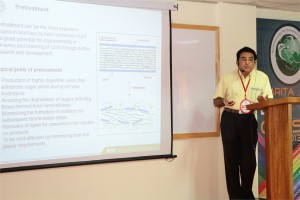
 Ashok Pandey, Ph.D.
Scientist F & Head, Biotechnology Division, National Institute for Interdisciplinary Science and Technology-CSIR), Thiruvananthapuram, India
Alternative renewable resources: Issues and perspectives for India – the case of transport fuels
With the increase in the urbanization way of life and also more and more dependence on materialistic life, there is substantial growing demand for the energy. The science and technological policy of the India has looked several avenues to fulfill this demand through alternative resources such as solar energy, wind energy, tidal energy, bioenergy, etc. The demand for the transport sector is largely met through the import (~70%). Biofuels, in particular bioethanol from lignocellulosic biomass offer attractive possibilities in this regard.
The sugar platform which generates ethanol is considered to be the most valuable solution to the transport fuel demand. Bioethanol can be generated from grains as well as from lignocellulosic plant material by their saccharification to sugars and subsequent fermentation of the sugars to produce ethanol. Bio-ethanol as a transportation fuel is attractive since it is more energy efficient than gasoline and produces less emissions. The benefits of developing biomass to ethanol technology(s) include: increased national energy security, reduction in GHG emissions, use of renewable resources, economic benefits and creation of employment and the foundation of a carbohydrate based chemical industry. However, the utilization of lignocellulosic biomass for fuel generation has not been given the sort of attention it ought to receive. It is known that the technology for ethanol production from biomass has to evolve greatly for an economical commercial scale utilization of the renewable biomass resources. Biomass requires extensive processing involving multiple steps for hydrolysis and fermentation of the raw material for producing ethanol. Feed stock availability, pretreatment, saccharification, fermentation and ethanol recovery are all factors which influence the production of ethanol and which needs R&D efforts for overall improvement of the production economics.
Bioconversion of lignocellulosic biomass (LB) can contribute significantly to the production of organic chemicals also. LB is also considered to be the only foreseeable source of energy. LB is mainly composed of (dry wt basis): cellulose, 40-60; hemicellulose, 20-40; and lignin, 10-25%. Most efficient method of biomass hydrolysis is through enzymatic saccharification5 using cellulases and hemicellulases. Fungal cellulases (FCs) have proved to be a better candidate than other microbial cellulases, with their secreted free cellulase complexes comprising all three components of cellulase [endoglucanases, exoglucanases and cellobiases (glucosidases).
The Centre for Biofuels at NIIST, Trivandrum, India aims ultimately to develop technologies and processes which will address the nation’s need for making fuel ethanol from the renewable resource: biomass. It is proposed to direct R&D activities at the major requirements of a biomass-ethanol technology, which include production of cellulases, hydrolysis of biomass, and ethanol fermentation. Viable technologies for each of these processes will contribute to the overall process development for fuel alcohol production from cheap and renewable biomass resources.
The lecture would present perspectives on bioethanol from lignocellulosic feedstocks.
References
- Biofuels- Alternative Feedstocks and Conversion Processes, Editors- Ashok Pandey, C Larroche, SC Ricke, CG Dussap & E Gnansounou, Academic Press, Elsevier Inc; San Diego, USA, p629 (2011) ISBN: 978-0-12-385099-7
- Handbook of Plant-Based Biofuels, Editor- Ashok Pandey, CRC Press, Francis & Taylors, Boca Raton, USA, p 297 (2008) ISBN 978-q-5602-2175-3
- Biofuels II, Special issue of Journal of Scientific & Industrial Research, Guest Editors- E Gnansounou, C Larroche and Ashok Pandey, 67(11), 837-1040 (2008) ISSN: 0022-4456
- Biofuels, Special issue of Journal of Scientific & Industrial Research, Guest Editors- C Larroche and Ashok Pandey, 64(11), 797-988 (2005) ISSN: 0022-4456
 Claudia AM Wheeler-Kingshott, Ph.D.
University Reader in Magnetic Resonance Physics, Department of Neuroinflammation, UCL Institute of Neurology, London, UK
Abstract
Detecting neuronal activity in vivo non-invasively is possible with a number of techniques. Amongst these, in 1990 functional magnetic resonance imaging (fMRI) was proposed as a technique that has a great ability to spatially map brain activity by exploiting the blood oxygenation level dependent (BOLD) contrast mechanism [1, 2]. In fact, neuronal activation triggers a demand for oxygen and induces a localised increase in blood flow and blood volume, which actually exceeds the metabolic needs. This in turns causes an increase of oxyhaemoglobin in the venous compartment, which is a transient phenomenon and is accompanied by a transient change (decrease) in the concentration of deoxyhaemoglobin. Due to its paramagnetic properties, the amount of deoxyhaemoglobin present in the venous blood affects the local magnetic field seen by the spins (protons) and determines the local properties of the MR signal. A decrease in deoxyhaemoglobin during neuronal activity, therefore, induces local variations of this magnetic field that increases the average transverse relaxation time of tissue, measured via the T2* parameter [3]. This means that there is an increase of the MR signal (of the order of a few %, typically <5%) linked to metabolic changes happening during brain function. Activation can be inferred at different brain locations by performing tasks while acquiring the MR signal and comparing periods of rest to periods of activity.
The macroscopic changes of the BOLD signal are well characterised, while the reason for the increased blood supply, exceeding demands, needs further thoughts. Here we will discuss two approaches for explaining the BOLD phenomenon, one that links it to adenosine triphosphate production [4] and enzyme saturation, the other that relates it to the very slow diffusion of oxygen through the blood-brain-barrier with a consequent compensatory high demand of oxygen [5]. Some evidence of restricted oxygen diffusion has been shown by means of hypercapnia [6], although it is not excluded that both mechanisms may be present.
Overall, the BOLD signal changes theory and its physiological basis will be presented and discussed.
References
- Ogawa, S., et al., Brain magnetic resonance imaging with contrast dependent on blood oxygenation. Proc Natl Acad Sci U S A, 1990. 87(24): p. 9868-72.
- Kwong, K.K., et al., Dynamic magnetic resonance imaging of human brain activity during primary sensory stimulation. Proc Natl Acad Sci U S A, 1992. 89(12): p. 5675-9.
- Bandettini PA, et al. Spin-echo and gradient-echo EPI of human brain activation using BOLD contrast: a comparative study at 1.5 T. NMR Biomed. 1994 Mar;7(1-2):12-20
- Fox, P.T., et al., Nonoxidative glucose consumption during focal physiologic neural activity. Science, 1988. 241(4864): p. 462-4.
- Gjedde, A., et al. Reduction of functional capillary density in human brain after stroke. J Cereb Blood Flow Metab, 1990. 10(3): p. 317-26.
- Hoge, R.D., et al., Linear coupling between cerebral blood flow and oxygen consumption in activated human cortex. Proc Natl Acad Sci U S A, 1999. 96(16): p. 9403-8.
 K. Satyamoorthy, Ph.D.
Director, Life Sciences Centre, Manipal University, India
Epigenetic Changes due to DNA Methylation in Human Epithelial Tumors
Extensive global hypomethylation in the genome and hypermthylation of selective tumor specific suppressor genes appears to be a hallmark of human cancers. Data suggests that hypermethylation of promoter region in genes is more closely related to subsequent gene expression; contrary to gene-body DNA methylation. The intricate balance between these two may contribute to the progressive process of development, differentiation and carcinogenesis. Epigenetic changes encompass, apart from DNA methylation, chromatin modifications through post-translational changes in histones and control by miRNAs. At the genome level, effects from these are compounded by copy number variations (CNVs) which may ultimately influence protein functions. From clinical perspective, changes in DNA methylation occur very early which are reversible and are influenced by environmental factors. Therefore, these can be potential resource for identifying therapeutic targets as well as biomarkers for early screening of cancer. Our current efforts in profiling genome wide DNA methylation changes in oral, cervical and breast cancers through DNA methylation microarray analysis has revealed number of alterations critical for survival, progression and metastatic behavior of tumors. Bioinformatics and functional analysis revealed several key regulatory molecules controlled by DNA methylation and suggests that DNA methylation changes in several CpG islands appear to co-segregate in the regions of miRNAs as well as in the CNVs. We have validated the signatures for methylation of CpG islands through bisufite sequencing for essential genes in clinical samples and have undertaken transcriptional and functional analysis in tumor cell lines. These results will be presented.
 Ajith Madhavan
Assistant Professor, School of Biotechnology, Amrita University
Development of a Phototrophic Microbial Fuel Cell with sacrificial electrodes and a novel proton exchange matrix
If micro organisms can solve Sudoku and possibly have feelings, who is to say that they cannot also solve the planet’s energy crisis? Mr. Madhavan employs micro organisms to produce energy using microbial fuel cell (MFC). Micro organisms go through a series of cycles and pathways in order to survive, including the Electron Transport Pathway (ETP) in which bacteria release electrons which can be tapped as energy. In a two-chambered MFC, micro organisms interact with an anode in one chamber and in the presence of an oxidizing agent in the cathodic chamber scavenges electrons from the cathode. The two chambers are connected by an external circuit and connected to a load. In between the two chambers is a proton exchange membrane (PEM) which transports protons from the second chamber to the first and acts as a barrier for electrons. Therefore, a renewable source of energy can be maintained by just providing your bacterial culture with the proper nutrients to thrive and remain happy and satisfied (assuming they have emotions).
Mr. Madhavan has done extensive work on such MFCs and has experimented with various micro organisms and substrates to achieve high energy production. The phototropic MFC Mr. Madhavan designed using Synechococcus elongates using waste water as a substrate was able to generate approximately 10 mȦ and 1 volt of electricity. Other research in this area has even shown that using human urine can be used as a substrate for certain bacteria to produce enough energy to charge a mobile phone.
Although this microbial technology seems to be the “next big thing” (despite their small size) when it comes to renewable energy sources there is still a lot of work to be done before these bacteria batteries hit the market. As of now the MFCs are still much less efficient than solar cells and the search for the perfect bacteria and substrate continues.

Abhijeet Kate, Arpana G Panicker, Diana Writer, Giridharan P, Keshav K V Ramamoorthy, Saji George, Shailendra K Sonawane
Protoplast fusion and transformation: A tool for activation of latent gene clusters
In the quest to discover new bioactive leads for unmet medical needs, actinomycetes present a treasure trove of undiscovered molecules. The ability of actinomycetes to produce antibiotics and other bioactive secondary metabolites has been underestimated due to sparse studies of cryptic gene clusters. These gene clusters can be tapped to explore scaffolds hidden in them. The up-regulation of the dormant genes is one of the most important areas of interest in the bioactive compounds discovery from microbial resources. Genome shuffling is a powerful tool for the activation of such gene clusters. Lei Yu, et al.1, reported enhancement of the lactic acid production in Lactobacillus rhamnosus through genome shuffling brought about by protoplast fusion. D. A. Hopwood et al.2 suggested that an interspecific recombination between strains producing different secondary metabolites, generate producers of ‘hybrid’ antibiotics. They also mentioned that an intraspecific fusion of actinomycetes protoplast bring about random and high frequency recombination. Protoplasts can also be used as recipients for isolated DNA, again in the presence of polyethylene glycol (PEG). In our study we had undertaken random genome shuffling by protoplast fusion of two, rather poorly expressed actinomycetes strains A (Figure 1) & B (Figure 2), mediated by PEG; and also by naked DNA transformation of Strain A protoplast with the DNA of Strain B. We generated eight protoplast fusants and seven transformants from parents considering their morphological difference from the two parent strains. These 15 recombinants were checked for their same colony morphologies for five generations to ensure phenotypic stability. Antibiotic resistance pattern was established by using antibiotic octodisc to generate a marker profile of the recombinants and the parent strains. Eight fusants (AP-18, AP-25, AP-2, AP-11, AP-14, AP-19, AP-11 and AP-27) and four transformants (TAP-30, TAP-31, TAP-32 and TAP-33) (Table 1) have shown a different antibiotic sensitivity pattern as compared to the parent strains. We envisage that these recombinants harbor shuffled gene clusters. To support array of conditions to express such shuffled/cryptic genes the recombinants were fermented in 11 different nutrient stress variants. The extracts generated were subjected to metabolite profiling by HPLC-ELSD, bioactivity screening for cytotoxicity and anti-infective capabilities. Two fusants AP-11 (Figure 3) and AP-25; one transformant TAP-32 (in growth media MBA-5 and MBA-7) displayed antifungal activity unlike parent strains (Table 2) Fusant AP-11 (Table 5) exhibited significant cell growth inhibition of five different cancer cell lines. The parents Strain A and Strain B did not exhibit any cell growth inhibition of these cell lines (Table 5). The metabolite profiling of fusant AP-11 and transformant TAP-32 was done by HPLC-ELSD. AP-11 showed the presence of five additional peaks (Figure 5 & Figure 6); TAP-32 extract from medium MBA-5 (Figure 7 & Figure 8) showed the presence of four additional peaks and TAP-32 extract from MBA-7 (Figure 9 & Figure 10) showed 14 additional peaks as compared to parent strains in similar medium and media controls. The study indicated that protoplast fusion and transformation have not only caused morphological changes but also shuffled genes responsible for synthesis of bioactive molecules. Further characterization of these new peaks is warranted.
 Shrikant Anant, Ph.D.
The Department of Molecular & Integrative Physiology, Kansas University Medical Center, USA
Cancer Stem Cells: Target Colon Cancers
Shrikant Anant, Deep Kwatra and Dharmalingam Subramaniam
Colon cancer is a leading cause of cancer related deaths in the US, and its rate is increasing at an alarming rate in lndia. Recent studies have suggested the drug resistance role for a mall number of cells within a tumor called cancer stem cells. We identified the colon cancer stem cell marker DCLK1, a member of the protein kinase superfamily and the doublecortin family. The protein encodes a Cterminal serinethreonine protein kinase domain, which shows substantial homology to Ca2calmodulindependent protein kinase. Our current studies have been to identify compounds that can either affect DCLK1 expression or inhibits its activity as a way to inhibit cancer stem cells. Honokiol is a biphenolic compound that has been used in the traditional Chinese Medicine for treating various ailments. In vitro kinase assays with recombinant DCLK1 demonstrated that honokiol inhibits its kinase activity in a dose dependent manner. We therefore determined the effect of honokiol on stem cells. One method to look at effects on stem cells is perform a spheroid assay, where spheroids formation is suggested to maintain stemlike characteristic of cancer cells. Honokiol significantly suppressed colonosphere formation of two colon cancer cell lines HCT116 and SW480. Flow cytometry studies confirmed that honokiol reduced the number of DCLK1cells. A critical signaling pathway known to modulate intestinal stem cell proliferation is the Hippo signaling pathway, and deregulation of the pathway leads to tumor development. DCLK1cells had high levels of YAP1, the nuclear target of Hippo signaling. We determined the effect of honokiol on components of the hipposignaling pathway. Honokiol reduced the phosphorylation of Mst1/2, Lats1/2 and YAP1. Furthermore, honokiol treatment resulted in downregulation of YAPTEAD complex protein TEAD-1. Ectopic expression of the TEAD-1 partially rescued the cells from honokiol mediated growth suppression. To determine the effect of honokiol on tumor growth in vivo, nude mice harboring HCT116 tumor xenografts in their flanks were administered the compound intraperitoneally every day for 21 days. Honokiol treatment significantly inhibited tumor xenograft growth. Western blot and immunohistochemistry analyses demonstrated significant inhibition in the expression of stem marker and Hippo signaling proteins in the honokioltreated xenograft tissues. Taken together, these data suggest that honokiol is a potent inhibitor of colon cancer that targets DCLK1 stem cells by inhibiting Hippo signaling pathway.

