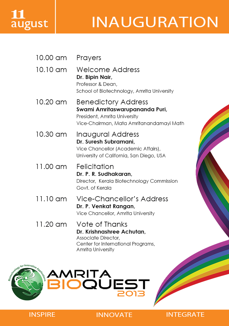


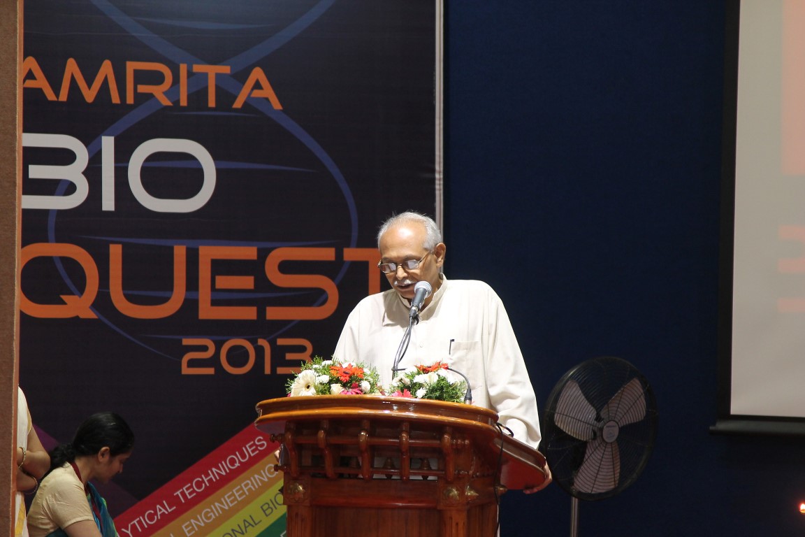


 Leland H. Hartwell Ph.D.
2001 Nobel Laureate, Physiology & Medicine
Dr. Lee Hartwell received the 2001 Nobel Prize in Physiology / Medicine for his discovery of protein molecules that control the division of cells. He was the President and Director of the Fred Hutchinson Cancer Research Center in Seattle, Washington before moving to Arizona State University’s Center for Sustainable Health.
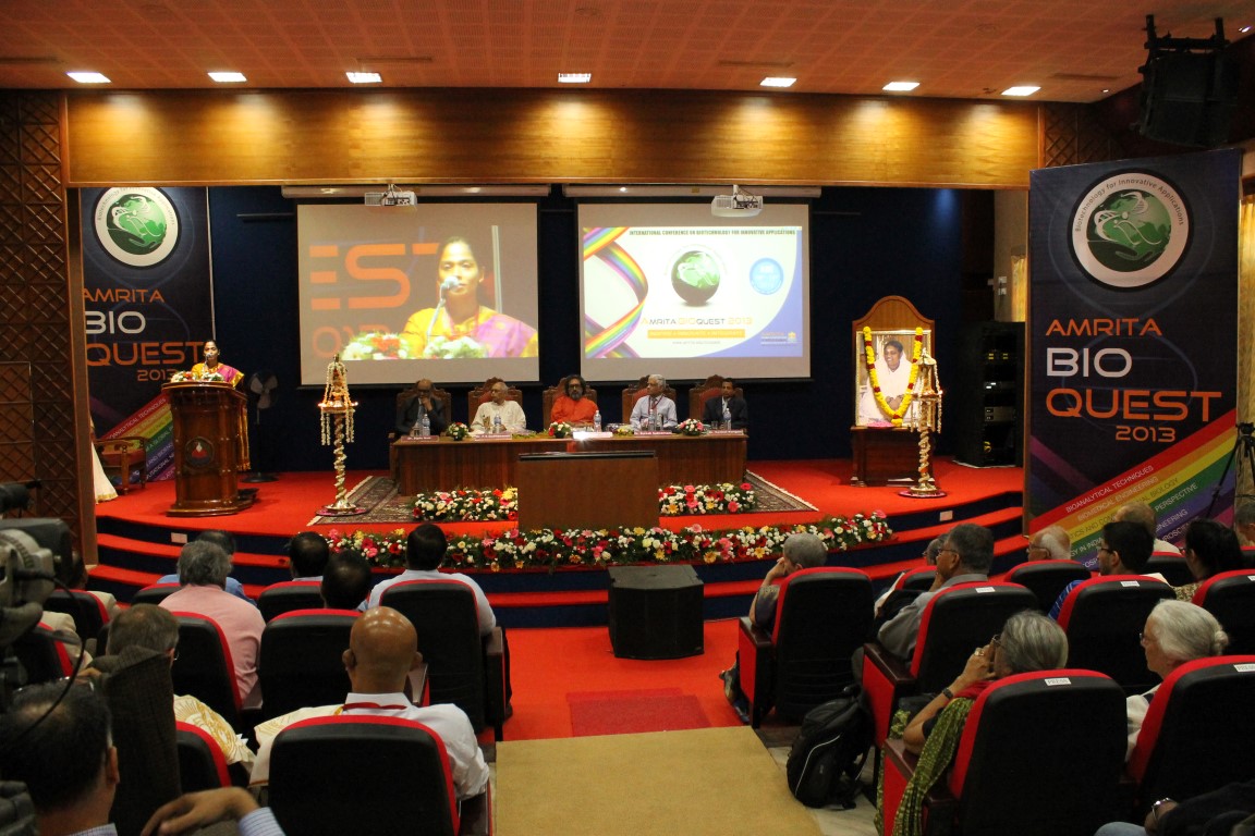
Dr. Hartwell is also adjunct faculty at Amrita University. He spoke to the delegates at Bioquest from his office in the US, over Amrita’s e-learning platform A-View. Given below are excerpts from his address.
I would like to address the young people in the audience. I know that many of you may have come to this meeting wondering, “How can I become a successful scientist? How can I prepare myself to make a contribution in this world?”
These questions are interesting to me also.
Believe it or not, I am still trying to be a successful scientist. That may surprise you since you probably think that a Nobel laureate must have found the answers. But the problem is that the answers to these questions change with time and the answers are different today than what they were when I began my career fifty years ago. The strategy of the 1960’s doesn’t work so well anymore. What is different now?
First, what we know now is much more. For example, by 1970, no genes from any organisms were sequenced. In 2013, we have the complete sequence of the human genome. Second, not only do we know much more today, accessing that knowledge is easy. Third, obtaining new information is much faster today.
Our rich understanding of science and technology is now needed to solve many serious problems. The human population has reached the size where we are utilizing all available resource of the planet. We are utilizing all of the agricultural land, all of the water, all of the forest and fishing resources. We are also polluting the planet that we live on.
We are polluting the land with fertilizers and pesticides; the oceans with acids and the atmosphere with carbon dioxide. We are using up top soil and ground water, thereby reducing our capacity to feed ourselves. We are using up petroleum, the energy source that our entire economy is dependent on. These are problems we were largely unaware of, fifty years ago. But these are problems that must be solved in your life times.
The big question facing your generation is, how can human beings live sustainably on planet earth. Your two broad goals on sustainability are 1) leave the planet as you first found it for your future generations; don’t use up the resources and don’t pollute the planet 2) everyone deserves to have an equal share of the earth’s resources.
Income strongly determines one’s opportunities in life. Many poor people succumb to chronic diseases and unhealthy environments. This inequality undermines our ability to live sustainably. We can’t ask the poor to leave the planet as they found it if they can’t support their families. Education, healthcare, employment are essential to having a sustainable society.
How can we be a successful scientist in 2013?
1. First choose a problem to solve
2. Ask questions to understand why it is not solved
3. Collaborate with those who can help
4. Develop a solution that works in the real worldChronic diseases are our major burden and this burden will get worse. Heart disease, diabetes, cancer, dementia and other diseases. The good news is that the chronic diseases are largely preventable and more easily curable if detected early. One question that attracts me is how can we detect disease earlier when it can be more easily cured?
Can we use our increasing knowledge in molecular biology to identify biomarkers for early disease detection?
We need to collaborate very closely with clinicians who care for patients to find out exactly where they need help.
I think if we apply our technology to important clinical questions we will actually save medical expenditure and be well on our way to making a great contribution to society.


Aswath Balakrishnan, Kapaettu Satyamoorthy and Manjunath B Joshi
Introduction
Insulin resistance is a hall mark of metabolic disorders such as diabetes. Reduced insulin response in vasculature leads to disruption of IR/Akt/eNOS signaling pathway resulting in vasoconstriction and subsequently to cardiovascular diseases. Recent studies have demonstrated that inflammatory regulator interleukin-6 (IL-6), as one of the potential mediators that can link chronic inflammation with insulin resistance. Accumulating evidences suggest a significant role of epigenetic mechanisms such as DNA methylation in progression of metabolic disorders. Hence the present study aimed to understand the role of epigenetic mechanisms involved during IL-6 induced vascular insulin resistance and its consequences in cardiovascular diseases.
Materials and Methods
Human umbilical vein endothelial cells (HUVEC) and Human dermal microvascular endothelial cells (HDMEC) were used for this study. Endothelial cells were treated in presence or absence of IL-6 (20ng/ml) for 36 hours and followed by insulin (100nM) stimulation for 15 minutes. Using immunoblotting, cell lysates were stained for phosphor- and total Akt levels to measure insulin resistance. To investigate changes in DNA methylation, cells were treated with or without neutrophil conditioned medium (NCM) as a physiological source of inflammation or IL-6 (at various concentrations) for 36 hours. Genomic DNA was processed for HPLC analysis for methyl cytosine content and cell lysates were analyzed for DNMT1 (DNA (cytosine-5)-methyltransferase 1) and DNMT3A (DNA (cytosine-5)-methyltransferase 3A) levels using immunoblotting.
Results
Endothelial cells stimulated with insulin exhibited an increase in phosphorylation of Aktser 473 in serum free conditions but such insulin response was not observed in cells treated with IL-6, suggesting chronic exposure of endothelial cells to IL-6 leads to insulin resistance. HPLC analysis for global DNA methylation resulted in decreased levels of 5-methyl cytosine in cells treated with pro-inflammatory molecules (both by NCM and IL-6) as compared to untreated controls. Subsequently, analysis in cells treated with IL-6 showed a significant decrease in DNMT1 levels but not in DNMT3A. Other pro-inflammatory marker such as TNF-α did not exhibit such changes.
Conclusion
Our study suggests: a) Chronic treatment of endothelial cells with IL-6 results in insulin resistance b) Neutrophil conditioned medium and IL-6 decreases methyl cytosine levels c) DNMT1 but not DNMT3a levels are reduced in cells treated with IL-6.
 Sudarslal S, Ph.D.
Associate Professor, School of Biotechnology, Amrita University
Electrospray ionization ion trap mass spectrometry for cyclic peptide characterization
There has been considerable interest in the isolation and structural characterization of bioactive peptides produced by bacteria and fungi. Most of the peptides are cyclic depsipeptides characterized by the presence of lactone linkages and β-hydroxy fatty acids. Occurrence of microheterogeneity is another remarkable property of these peptides. Even if tandem mass spectrometers are good analytical tools to structurally characterize peptides and proteins, sequence analysis of cyclic peptides is often ambiguous due to the random ring opening of the peptides and subsequent generation of a set of linear precursor ions with the same m/z. Here we report combined use of chemical derivatization and multistage fragmentation capability of ion trap mass spectrometers to determine primary sequences of a series of closely related cyclic peptides.


Ravindra Gudihal, Suresh Babu C V
Bioanalytical Characterization of Therapeutic Proteins
The characterization of therapeutic proteins such as monoclonal antibody (mAb) during different stages of manufacturing is crucial for timely and successful product release. Regulatory agencies require a variety of analytical technologies for comprehensive and efficient protein analysis. Electrophoresis-based techniques and liquid chromatography (LC) either standalone or coupled to mass spectrometry (MS) are at the forefront for the in-depth analysis of protein purity, isoforms, stability, aggregation, posttranslational modifications, PEGylation, etc. In this presentation, a combination of various chromatographic and electrophoretic techniques such as liquid-phase isoelectric focusing, microfluidic and capillary-based electrophoresis (CE), liquid chromatography (LC) and combinations of those with mass spectrometry techniques will be discussed. We present a workflow based approach to the analysis of therapeutic proteins. In successive steps critical parameters like purity, accurate mass, aggregation, peptide sequence, glycopeptide and glycan analysis are analyzed. In brief, the workflow involved proteolytic digestion of therapeutic protein for peptide mapping, N-Glycanase and chemical labeling reaction for glycan analysis, liquid-phase isoelectric focusing for enrichment of charge variants followed by a very detailed analysis using state of the art methods such as CE-MS and LC-MS. For the analysis of glycans, we use combinations of CE-MS and LC-MS to highlight the sweet spots of these techniques. CE-MS is found to be more useful in analysis of highly sialylated glycans (charged glycans) while nano LC-MS seems to be better adapted for analysis of neutral glycans. These two techniques can be used to get complementary data to profile all the glycans present in a given protein. In addition, microfluidic electrophoresis was used as a QC tool in initial screening for product purity, analysis of papain digestion fragments of mAb, protein PEGylation products, etc. The described workflow involves multiple platforms, provides an end to end solution for comprehensive protein characterization and aims at reducing the total product development time.