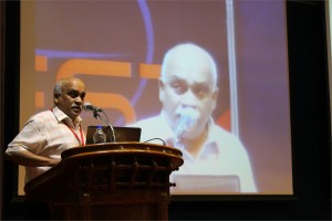
 Gaute Einevoll, Ph.D.
Professor of Physics, Department of Mathematical Sciences & Technology, Norwegian University of Life Sciences (UMB)
Multiscale modeling of cortical network activity: Key challenges
Gaute T. Einevoll Computational Neuroscience Group, Norwegian University of Life Sciences, 1432 Ås, Norway; Norwegian National Node of the International Neuroinformatics Coordinating Facility (INCF)
Several challenges must be met in the development of multiscale models of cortical network activity. In the presentation I will, based on ongoing work in our group (http://compneuro.umb.no/ ) on multiscale modeling of cortical columns, outline some of them. In particular,
-
Combined modeling schemes for neuronal, glial and vascular dynamics [1,2],
-
Development of sets of interconnected models describing system at different levels of biophysical detail [3-5],
-
Multimodal modeling, i.e., how to model what you can measure [6-12],
-
How to model when you don’t know all the parameters, and
-
Development of neuroinformatics tools and routines to do simulations efficiently and accurately [13,14].
References:
- L. Øyehaug, I. Østby, C. Lloyd, S.W. Omholt, and G.T. Einevoll: Dependence of spontaneous neuronal firing and depolarisation block on astroglial membrane transport mechanisms, J Comput Neurosci 32, 147-165 (2012)
- I. Østby, L. Øyehaug, G.T. Einevoll, E. Nagelhus, E. Plahte, T. Zeuthen, C. Lloyd, O.P. Ottersen, and S.W. Omholt: Astrocytic mechanisms explaining neural-activity-induced shrinkage of extraneuronal space, PLoS Comp Biol 5, e1000272 (2009)
- T. Heiberg, B. Kriener, T. Tetzlaff, A. Casti, G.T. Einevoll, and H.E. Plesser: Firing-rate models can describe the dynamics of the retina-LGN connection, J Comput Neurosci (2013)
- T. Tetzlaff, M. Helias, G.T. Einevoll, and M. Diesmann: Decorrelation of neural-network activity by inhibitory feedback, PLoS Comp Biol 8, e10002596 (2012).
- E. Nordlie, T. Tetzlaff, and G.T. Einevoll: Rate dynamics of leaky integrate-and-fire neurons with strong synapses, Frontiers in Comput Neurosci 4, 149 (2010)
- G.T. Einevoll, F. Franke, E. Hagen, C. Pouzat, K.D. Harris: Towards reliable spike-train recording from thousands of neurons with multielectrodes, Current Opinion in Neurobiology 22, 11-17 (2012)
- H. Linden, T Tetzlaff, TC Potjans, KH Pettersen, S Grun, M Diesmann, GT Einevoll: Modeling the spatial reach of LFP, Neuron 72, 859-872 (2011).
- H. Linden, K.H. Pettersen, and G.T. Einevoll: Intrinsic dendritic filtering gives low-pass power spectra of local field potentials, J Computational Neurosci 29, 423-444 (2010)
- K.H. Pettersen and G.T. Einevoll: Amplitude variability and extracellular low-pass filtering of neuronal spikes, Biophysical Journal 94, 784-802 (2008).
- K.H. Pettersen, E. Hagen, and G.T. Einevoll: Estimation of population firing rates and current source densities from laminar electrode recordings, J Comput Neurosci 24, 291-313 (2008).
- K. Pettersen, A. Devor, I. Ulbert, A.M. Dale and G.T. Einevoll. Current-source density estimation based on inversion of electrostatic forward solution: Effects of finite extent of neuronal activity and conductivity discontinuities, Journal of Neuroscience Methods 154, 116-133 (2006).
- G.T. Einevoll, K. Pettersen, A. Devor, I. Ulbert, E. Halgren and A.M. Dale: Laminar Population Analysis: Estimating firing rates and evoked synaptic activity from multielectrode recordings in rat barrel cortex, Journal of Neurophysiology 97, 2174-2190 (2007).
- LFPy: A tool for simulation of extracellular potentials (http://compneuro.umb.no)
- E. Nordlie, M.-O. Gewaltig, H. E. Plesser: Towards reproducible descriptions of neuronal network models, PLoS Comp Biol 5, e1000456 (2009).
 Claudia AM Wheeler-Kingshott, Ph.D.
University Reader in Magnetic Resonance Physics, Department of Neuroinflammation, UCL Institute of Neurology, London, UK
Abstract
Detecting neuronal activity in vivo non-invasively is possible with a number of techniques. Amongst these, in 1990 functional magnetic resonance imaging (fMRI) was proposed as a technique that has a great ability to spatially map brain activity by exploiting the blood oxygenation level dependent (BOLD) contrast mechanism [1, 2]. In fact, neuronal activation triggers a demand for oxygen and induces a localised increase in blood flow and blood volume, which actually exceeds the metabolic needs. This in turns causes an increase of oxyhaemoglobin in the venous compartment, which is a transient phenomenon and is accompanied by a transient change (decrease) in the concentration of deoxyhaemoglobin. Due to its paramagnetic properties, the amount of deoxyhaemoglobin present in the venous blood affects the local magnetic field seen by the spins (protons) and determines the local properties of the MR signal. A decrease in deoxyhaemoglobin during neuronal activity, therefore, induces local variations of this magnetic field that increases the average transverse relaxation time of tissue, measured via the T2* parameter [3]. This means that there is an increase of the MR signal (of the order of a few %, typically <5%) linked to metabolic changes happening during brain function. Activation can be inferred at different brain locations by performing tasks while acquiring the MR signal and comparing periods of rest to periods of activity.
The macroscopic changes of the BOLD signal are well characterised, while the reason for the increased blood supply, exceeding demands, needs further thoughts. Here we will discuss two approaches for explaining the BOLD phenomenon, one that links it to adenosine triphosphate production [4] and enzyme saturation, the other that relates it to the very slow diffusion of oxygen through the blood-brain-barrier with a consequent compensatory high demand of oxygen [5]. Some evidence of restricted oxygen diffusion has been shown by means of hypercapnia [6], although it is not excluded that both mechanisms may be present.
Overall, the BOLD signal changes theory and its physiological basis will be presented and discussed.
References
- Ogawa, S., et al., Brain magnetic resonance imaging with contrast dependent on blood oxygenation. Proc Natl Acad Sci U S A, 1990. 87(24): p. 9868-72.
- Kwong, K.K., et al., Dynamic magnetic resonance imaging of human brain activity during primary sensory stimulation. Proc Natl Acad Sci U S A, 1992. 89(12): p. 5675-9.
- Bandettini PA, et al. Spin-echo and gradient-echo EPI of human brain activation using BOLD contrast: a comparative study at 1.5 T. NMR Biomed. 1994 Mar;7(1-2):12-20
- Fox, P.T., et al., Nonoxidative glucose consumption during focal physiologic neural activity. Science, 1988. 241(4864): p. 462-4.
- Gjedde, A., et al. Reduction of functional capillary density in human brain after stroke. J Cereb Blood Flow Metab, 1990. 10(3): p. 317-26.
- Hoge, R.D., et al., Linear coupling between cerebral blood flow and oxygen consumption in activated human cortex. Proc Natl Acad Sci U S A, 1999. 96(16): p. 9403-8.
 Shantikumar Nair, Ph.D.
Professor & Director, Amrita Center for Nanosciences & Molecular Medicine, Amrita University, India
Spatially Distributed and Hierarchical Nanomaterials in Biotechnology
Although nano materials are well investigated in biotechnology in their zero-, one- and two-dimensional forms, three-dimensional nanomaterials are relatively less investigated for their biological applications. Three dimensional nano materials are much more complex with several structural and hierarchical variables controlling their mechanical, chemical and biological functionality. In this talk examples are given of some complex three dimensional systems including, scaffolds, aggregates, fabrics and membranes. Essentially three types of hierarchies are considered: one-dimensional hierarchy, two-dimensional hierarchy and three-dimensional hierarchy each giving rise to unique behaviors.
 D. Narasimha Rao, Ph.D.
Professor, Dept of Biochemistry, Indian Institute of Science, Bangalore, India
Genomics of Restriction-Modification Systems
Restriction endonucleases occur ubiquitously among procaryotic organisms. Up to 1% of the genome of procaryotic organisms is taken up by the genes for these enzymes. Their principal biological function is the protection of the host genome against foreign DNA, in particular bacteriophage DNA. Restriction-modification (R-M) systems are composed of pairs of opposing enzyme activities: an endonuclease and a DNA methyltransferase (MTase). The endonucleases recognise specific sequences and catalyse cleavage of double-stranded DNA. The modification MTases catalyse the addition of a methyl group to one nucleotide in each strand of the recognition sequence using S-adenosyl-L-methionine (AdoMet) as the methyl group donor. Based on their molecular structure, sequence recognition, cleavage position and cofactor requirements, R-M systems are generally classified into three groups. In general R-M systems restrict unmodified DNA, but there are other systems that specifically recognise and cut modified DNA. More than 3500 restriction enzymes have been discovered so far. With the identification and sequencing of a number of R-M systems from bacterial genomes, an increasing number of these have been found that do not seem to fit into the conventional classification.
It is well documented that restriction enzyme genes always lie close to their cognate methyltransferase genes. Analysis of the bacterial and archaeal genome sequences shows that MTase genes are more common than one would have expected on the basis of previous biochemical screening. Frequently, they clearly form part of a R-M system, because the adjacent open reading frames (ORFs) show similarity to known restriction enzyme genes. Very often, though, the adjacent ORFs have no homologs in the GenBank and become candidates either for restriction enzymes with novel specificities or for new examples of previously uncloned specificities. Sequence-dependent modification and restriction forms the foundation of defense against foreign DNAs and thus RM systems may serve as a tool of defense for bacterial cells. RM systems however, sometimes behave as discrete units of life, and any threat to their maintenance, such as a challenge by a competing genetic element can lead to cell death through restriction breakage in the genome, thus providing these systems with a competitive advantage. The RM systems can behave as mobile-genetic elements and have undergone extensive horizontal transfer between genomes causing genome rearrangements. The capacity of RM systems to act as selfish, mobile genetic elements may underlie the structure and function of RM enzymes.
The similarities and differences in the different mechanisms used by restriction enzymes will be discussed. Although it is not clear whether the majority of R-M systems are required for the maintenance of the integrity of the genome or whether they are spreading as selfish genetic elements, they are key players in the “genomic metabolism” of procaryotic organisms. As such they deserve the attention of biologists in general. Finally, restriction enzymes are the work horses of molecular biology. Understanding their enzymology will be advantageous to those who use these enzymes, and essential for those who are devoted to the ambitious goal of changing the properties of these enzymes, and thereby make them even more useful.
 Satheesh Babu T. G., Ph.D.
Associate Professor, Department of Sciences, School of Engineering, Amrita University, Coimbatore, India
Nanomaterials for ‘enzyme-free’ biosensing
Enzyme based sensors have many draw backs such as poor storage stability, easily affected by the change in pH and temperature and involves complicated enzyme immobilization procedures. To address this limitation, an alternative approach without the use of enzyme, “non-enzymatic” has been tried recently. Choosing the right catalyst for direct electrochemical oxidation / reduction of a target molecule is the key step in the fabrication of non-enzymatic sensors.
Non-enzymatic sensors for glucose, creatinine, vitamins and cholesterol are fabricated using different nanomaterials, such as nanotubes, nanowires and nanoparticles of copper oxide, titanium dioxide, tantalum oxide, platinum, gold and graphenes. These sensors selectively catalyse the targeted analyte with very high sensitivity. These nanomaterials based sensors combat the drawbacks of enzymatic sensors.

Sunilkumar Sukumaran, Ayyappan Nair, Madhuri Subbiah, Gunja Gupta, Lakshmi Rajakrishna, Pradeep Savanoor Raghavendra, Subbulakshmi Karthikeyan, Salini Krishnan Unni and Ganesh Sambasivam
Genotoxicity is defined as DNA damage that leads to gene mutations which can become tumorigenic. Genotoxicity testing is important to ensure drug safety and is mandatory prior to Phase I/II clinical trials of new drugs. The results from genetic toxicology studies help to identify hazardous drugs and environmental genotoxins. Currently, among others there are four tests recommended by regulatory authorities (Ames test-bacterial, chromosome aberrations; in vitro gene mutation-eukaryotic cells and in vivo test). These assays are laborious, time consuming, require large quantities of test compounds and limited by throughput challenges. The site and mechanism of genotoxicity are not revealed by these assays and data obtained from bacterial tests might not translate the same in mammals. To address these we have developed a novel, versatile, human cell based, high throughput, reporter based genotoxicity screen (Anthem’s Genotox screen). This screen is performed on genetically engineered human cell lines that express 3 reporter genes under transcriptional control of ‘early DNA damage sensors’ (p53, p21 and GADD153). These genes are involved in DNA repair, cell cycle arrest and/or apoptosis. p21 and GADD are also known to be induced in a p53 independent manner. p53 blocks G1/S transition of cell cycle while the p53 independent DNA damage block G2/M transition. Identification of the mechanism of genotoxicity helps in rational drug designing. Additionally, the platform can be used to screen other potential genotoxins from cosmetics, food and environment. Initial validation studies of the Genotox screen was performed with over 60 compounds chosen from a variety of chemical classes. The genotoxic potential of metabolites was tested using rat liver S9 fractions. The results demonstrated a sensitivity of 86.7–92.3% and a specificity of 70–78.6% when compared with currently available in vitro genotoxicity assays. This Genotox screen would prove to be an invaluable human cell based tool to weed out potential genotoxins in various industries.


