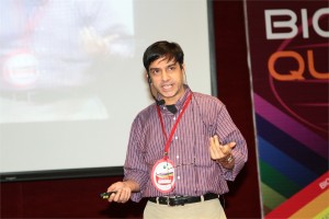
 Hideaki Nagase, Ph.D.
Kennedy Institute of Rheumatology-Centre for Degenerative Diseases, University of Oxford, UK
Osteoarthritis: diagnosis, treatment and challenges
Hideaki Nagase1, Ngee Han Lim1, George Bou-Gharios1, Ernst Meinjohanns2 and Morten Meldal3
- Kennedy Institute of Rheumatology, Nuffield Department of Orthopaedics, Rheumatology and Musculoskeletal Sciences, University of Oxford, London, W6 8LH UK
- Carlsberg Laboratory, Copenhagen, Denmark,
- Nano-Science Center, Department of Chemistry, University of Copenhagen, Denmark
Osteoarthritis (OA) is the most prevalent age-related degenerative joint disease. With the expanding ageing population, it imposes a major socio-economic burden on society. A key feature of OA is a gradual loss of articular cartilage and deformation of bone, resulting in the impairment of joint function. Currently, there is no effective disease-modifying treatment except joint replacement surgery. There are many possible causes of cartilage loss (e.g. mechanical load, injury, reactive oxygen species, aging, etc.) and etiological factors (obesity, genetics), but the degradation of cartilage is primarily caused by elevated levels of active metalloproteinases. It is therefore attractive to consider proteinase inhibitors as potential therapeutics. However, there are several hurdles to overcome, namely early diagnosis and continuous monitoring of the efficacy of inhibitor therapeutics. We are therefore aiming at developing non-invasive probes to detect cartilage degrading metalloproteinase activities.
We have designed in vivo imaging probes to detect MMP-13 (collagenase 3) activity that participates in OA by degrade cartilage collagen II and MMP-12 (macrophage elastase) activity involved in inflammatory arthritis. These activity-based probes consist of a peptide that is selectively cleaved by the target proteinase, a near-infrared fluorophore and a quencher. The probe’s signal multiplies upon proteolysis. They were first used to follow the respective enzyme activity in vivo in the mouse model of collagen-induced arthritis and we found MMP-12 activity probe (MMP12AP) activation peaked at 5 days after onset of the disease, whereas MMP13AP activation was observed at 10-15 days. The in vivo activation of these probes was inhibited by specific low molecule inhibitors. We proceeded to test both probes in the mouse model of OA induced by the surgical destabilization of medial meniscus of the knee joints. In this model, degradation of knee cartilage is first detected histologically 6 weeks after surgery with significant erosion detectable at 8 weeks. Little activation of MMP12AP was detected, which was expected, as macrophage migration is not obvious in OA. MMP13AP, on the other hand, was significantly activated in the operated knee at 6 weeks compared with the non-operated contralateral knee, but there were no significant differences between the operated and sham-operated knees. At 8 weeks, however, the signals in the operated knees were significantly higher than both the contralateral and sham-operated controls. Activation of aggrecanases and MMP-13 are observed before structural changes of cartilage. We are therefore currently improving the MMP-13 probe for earlier detection by attaching it to polymers that are retained in cartilage.
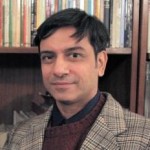 Rohit Manchanda, Ph.D.
Professor, Biomedical Engineering Group, IIT-Bombay, India
Modelling the syncytial organization and neural control of smooth muscle: insights into autonomic physiology and pharmacology
We have been studying computationally the syncytial organization and neural control of smooth muscle in order to help explain certain puzzling findings thrown up by experimental work. This relates in particular to electrical signals generated in smooth muscles, such as synaptic potentials and spikes, and how these are explicable only if three-dimensional syncytial biophysics are taken fully into account. In this talk, I shall provide an illustration of outcomes and insights gleaned from such an approach. I shall first describe our work on the mammalian vas deferens, in which an analysis of the effects of syncytial coupling led us to conclude that the experimental effects of a presumptive gap junction uncoupler, heptanol, on synaptic potentials were incompatible with gap junctional block and could best be explained by a heptanol-induced inhibition of neurotransmitter release, thus compelling a reinterpretation of the mechanism of action of this agent. I shall outline the various lines of evidence, based on indices of syncytial function, that we adduced in order to reach this conclusion. We have now moved on to our current focus on urinary bladder biophysics, where the questions we aim to address are to do with mechanisms of spike generation. Smooth muscle cells in the bladder exhibit spontaneous spiking and spikes occur in a variety of distinct shapes, making their generation problematic to explain. We believe that the variety in shapes may owe less to intrinsic differences in spike mechanism (i.e., in the complement of ion channels participating in spike production) and more to features imposed by syncytial biophysics. We focus especially on the modulation of spike shape in a 3-D coupled network by such factors as innervation pattern, propagation in a syncytium, electrically finite bundles within and between which the spikes spread, and some degree of pacemaker activity by a sub-population of the cells. I shall report two streams of work that we have done, and the tentative conclusions these have enabled us to reach: (a) using the NEURON environment, to construct the smooth muscle syncytium and endow it with synaptic drive, and (b) using signal-processing approaches, towards sorting and classifying the experimentally recorded spikes.
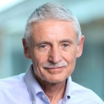 Leland H. Hartwell Ph.D.
2001 Nobel Laureate, Physiology & Medicine
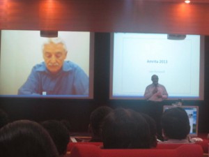
Dr. Lee Hartwell received the 2001 Nobel Prize in Physiology / Medicine for his discovery of protein molecules that control the division of cells. He was the President and Director of the Fred Hutchinson Cancer Research Center in Seattle, Washington before moving to Arizona State University’s Center for Sustainable Health.
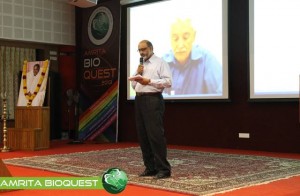
Dr. Hartwell is also adjunct faculty at Amrita University. He spoke to the delegates at Bioquest from his office in the US, over Amrita’s e-learning platform A-View. Given below are excerpts from his address.
I would like to address the young people in the audience. I know that many of you may have come to this meeting wondering, “How can I become a successful scientist? How can I prepare myself to make a contribution in this world?”
These questions are interesting to me also.
Believe it or not, I am still trying to be a successful scientist. That may surprise you since you probably think that a Nobel laureate must have found the answers. But the problem is that the answers to these questions change with time and the answers are different today than what they were when I began my career fifty years ago. The strategy of the 1960’s doesn’t work so well anymore. What is different now?
First, what we know now is much more. For example, by 1970, no genes from any organisms were sequenced. In 2013, we have the complete sequence of the human genome. Second, not only do we know much more today, accessing that knowledge is easy. Third, obtaining new information is much faster today.
Our rich understanding of science and technology is now needed to solve many serious problems. The human population has reached the size where we are utilizing all available resource of the planet. We are utilizing all of the agricultural land, all of the water, all of the forest and fishing resources. We are also polluting the planet that we live on.
We are polluting the land with fertilizers and pesticides; the oceans with acids and the atmosphere with carbon dioxide. We are using up top soil and ground water, thereby reducing our capacity to feed ourselves. We are using up petroleum, the energy source that our entire economy is dependent on. These are problems we were largely unaware of, fifty years ago. But these are problems that must be solved in your life times.
The big question facing your generation is, how can human beings live sustainably on planet earth. Your two broad goals on sustainability are 1) leave the planet as you first found it for your future generations; don’t use up the resources and don’t pollute the planet 2) everyone deserves to have an equal share of the earth’s resources.
Income strongly determines one’s opportunities in life. Many poor people succumb to chronic diseases and unhealthy environments. This inequality undermines our ability to live sustainably. We can’t ask the poor to leave the planet as they found it if they can’t support their families. Education, healthcare, employment are essential to having a sustainable society.
How can we be a successful scientist in 2013?
1. First choose a problem to solve
2. Ask questions to understand why it is not solved
3. Collaborate with those who can help
4. Develop a solution that works in the real worldChronic diseases are our major burden and this burden will get worse. Heart disease, diabetes, cancer, dementia and other diseases. The good news is that the chronic diseases are largely preventable and more easily curable if detected early. One question that attracts me is how can we detect disease earlier when it can be more easily cured?
Can we use our increasing knowledge in molecular biology to identify biomarkers for early disease detection?
We need to collaborate very closely with clinicians who care for patients to find out exactly where they need help.
I think if we apply our technology to important clinical questions we will actually save medical expenditure and be well on our way to making a great contribution to society.
 Shantikumar Nair, Ph.D.
Professor & Director, Amrita Center for Nanosciences & Molecular Medicine, Amrita University, India
Spatially Distributed and Hierarchical Nanomaterials in Biotechnology
Although nano materials are well investigated in biotechnology in their zero-, one- and two-dimensional forms, three-dimensional nanomaterials are relatively less investigated for their biological applications. Three dimensional nano materials are much more complex with several structural and hierarchical variables controlling their mechanical, chemical and biological functionality. In this talk examples are given of some complex three dimensional systems including, scaffolds, aggregates, fabrics and membranes. Essentially three types of hierarchies are considered: one-dimensional hierarchy, two-dimensional hierarchy and three-dimensional hierarchy each giving rise to unique behaviors.
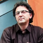 Nader Pourmand, Ph.D.
Director, UCSC Genome Technology Center,University of California, Santa Cruz
Biosensor and Single Cell Manipulation using Nanopipettes
Approaching sub-cellular biological problems from an engineering perspective begs for the incorporation of electronic readouts. With their high sensitivity and low invasiveness, nanotechnology-based tools hold great promise for biochemical sensing and single-cell manipulation. During my talk I will discuss the incorporation of electrical measurements into nanopipette technology and present results showing the rapid and reversible response of these subcellular sensors to different analytes such as antigens, ions and carbohydrates. In addition, I will present the development of a single-cell manipulation platform that uses a nanopipette in a scanning ion-conductive microscopy technique. We use this newly developed technology to position the nanopipette with nanoscale precision, and to inject and/or aspirate a minute amount of material to and from individual cells or organelle without comprising cell viability. Furthermore, if time permits, I will show our strategy for a new, single-cell DNA/ RNA sequencing technology that will potentially use nanopipette technology to analyze the minute amount of aspirated cellular material.
 Satheesh Babu T. G., Ph.D.
Associate Professor, Department of Sciences, School of Engineering, Amrita University, Coimbatore, India
Nanomaterials for ‘enzyme-free’ biosensing
Enzyme based sensors have many draw backs such as poor storage stability, easily affected by the change in pH and temperature and involves complicated enzyme immobilization procedures. To address this limitation, an alternative approach without the use of enzyme, “non-enzymatic” has been tried recently. Choosing the right catalyst for direct electrochemical oxidation / reduction of a target molecule is the key step in the fabrication of non-enzymatic sensors.
Non-enzymatic sensors for glucose, creatinine, vitamins and cholesterol are fabricated using different nanomaterials, such as nanotubes, nanowires and nanoparticles of copper oxide, titanium dioxide, tantalum oxide, platinum, gold and graphenes. These sensors selectively catalyse the targeted analyte with very high sensitivity. These nanomaterials based sensors combat the drawbacks of enzymatic sensors.
 Srisairam Achuthan, Ph.D.
Senior Scientific Programmer, Research Informatics Division, Department of Information Sciences, City of Hope, CA, USA
Applying Machine learning for Automated Identification of Patient Cohorts
Srisairam Achuthan, Mike Chang, Ajay Shah, Joyce Niland
Patient cohorts for a clinical study are typically identified based on specific selection criteria. In most cases considerable time and effort are spent in finding the most relevant criteria that could potentially lead to a successful study. For complex diseases, this process can be more difficult and error prone since relevant features may not be easily identifiable. Additionally, the information captured in clinical notes is in non-coded text format. Our goal is to discover patterns within the coded and non-coded fields and thereby reveal complex relationships between clinical characteristics across different patients that would be difficult to accomplish manually. Towards this, we have applied machine learning techniques such as artificial neural networks and decision trees to determine patients sharing similar characteristics from available medical records. For this proof of concept study, we used coded and non-coded (i.e., clinical notes) patient data from a clinical database. Coded clinical information such as diagnoses, labs, medications and demographics recorded within the database were pooled together with non-coded information from clinical notes including, smoking status, life style (active / inactive) status derived from clinical notes. The non-coded textual information was identified and interpreted using a Natural Language Processing (NLP) tool I2E from Linguamatics.

