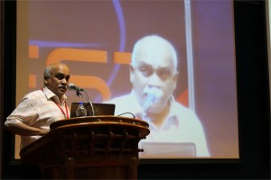
 Terry Hermiston, Ph.D.
Vice President, US Biologics Research Site Head, US Innovation Center Bayer Healthcare, USA
ColoAd1 – An oncolytic adenovirus derived by directed evolution
Attempts at developing oncolytic viruses have been primarily based on rational design. However, this approach has been met with limited success. An alternative approach employs directed evolution as a means of producing highly selective and potent anticancer viruses. In this method, viruses are grown under conditions that enrich and maximize viral diversity and then passaged under conditions meant to mimic those encountered in the human cancer microenvironment. Using the “Directed Evolution” methodology, we have generated ColoAd1, a novel chimeric oncolytic adenovirus. In vitro, this virus demonstrated a >2 log increase in both potency and selectivity when compared to ONYX-015 on colon cancer cells. These results were further supported by in vivo and ex vivo studies. Importantly, these results have validated this methodology as a new general approach for deriving clinically-relevant, highly potent anti-cancer virotherapies. This virus is currently in clinical trials as a novel treatment for cancer.

Abhijeet Kate, Arpana G Panicker, Diana Writer, Giridharan P, Keshav K V Ramamoorthy, Saji George, Shailendra K Sonawane
Protoplast fusion and transformation: A tool for activation of latent gene clusters
In the quest to discover new bioactive leads for unmet medical needs, actinomycetes present a treasure trove of undiscovered molecules. The ability of actinomycetes to produce antibiotics and other bioactive secondary metabolites has been underestimated due to sparse studies of cryptic gene clusters. These gene clusters can be tapped to explore scaffolds hidden in them. The up-regulation of the dormant genes is one of the most important areas of interest in the bioactive compounds discovery from microbial resources. Genome shuffling is a powerful tool for the activation of such gene clusters. Lei Yu, et al.1, reported enhancement of the lactic acid production in Lactobacillus rhamnosus through genome shuffling brought about by protoplast fusion. D. A. Hopwood et al.2 suggested that an interspecific recombination between strains producing different secondary metabolites, generate producers of ‘hybrid’ antibiotics. They also mentioned that an intraspecific fusion of actinomycetes protoplast bring about random and high frequency recombination. Protoplasts can also be used as recipients for isolated DNA, again in the presence of polyethylene glycol (PEG). In our study we had undertaken random genome shuffling by protoplast fusion of two, rather poorly expressed actinomycetes strains A (Figure 1) & B (Figure 2), mediated by PEG; and also by naked DNA transformation of Strain A protoplast with the DNA of Strain B. We generated eight protoplast fusants and seven transformants from parents considering their morphological difference from the two parent strains. These 15 recombinants were checked for their same colony morphologies for five generations to ensure phenotypic stability. Antibiotic resistance pattern was established by using antibiotic octodisc to generate a marker profile of the recombinants and the parent strains. Eight fusants (AP-18, AP-25, AP-2, AP-11, AP-14, AP-19, AP-11 and AP-27) and four transformants (TAP-30, TAP-31, TAP-32 and TAP-33) (Table 1) have shown a different antibiotic sensitivity pattern as compared to the parent strains. We envisage that these recombinants harbor shuffled gene clusters. To support array of conditions to express such shuffled/cryptic genes the recombinants were fermented in 11 different nutrient stress variants. The extracts generated were subjected to metabolite profiling by HPLC-ELSD, bioactivity screening for cytotoxicity and anti-infective capabilities. Two fusants AP-11 (Figure 3) and AP-25; one transformant TAP-32 (in growth media MBA-5 and MBA-7) displayed antifungal activity unlike parent strains (Table 2) Fusant AP-11 (Table 5) exhibited significant cell growth inhibition of five different cancer cell lines. The parents Strain A and Strain B did not exhibit any cell growth inhibition of these cell lines (Table 5). The metabolite profiling of fusant AP-11 and transformant TAP-32 was done by HPLC-ELSD. AP-11 showed the presence of five additional peaks (Figure 5 & Figure 6); TAP-32 extract from medium MBA-5 (Figure 7 & Figure 8) showed the presence of four additional peaks and TAP-32 extract from MBA-7 (Figure 9 & Figure 10) showed 14 additional peaks as compared to parent strains in similar medium and media controls. The study indicated that protoplast fusion and transformation have not only caused morphological changes but also shuffled genes responsible for synthesis of bioactive molecules. Further characterization of these new peaks is warranted.
 Nader Pourmand, Ph.D.
Director, UCSC Genome Technology Center,University of California, Santa Cruz
Biosensor and Single Cell Manipulation using Nanopipettes
Approaching sub-cellular biological problems from an engineering perspective begs for the incorporation of electronic readouts. With their high sensitivity and low invasiveness, nanotechnology-based tools hold great promise for biochemical sensing and single-cell manipulation. During my talk I will discuss the incorporation of electrical measurements into nanopipette technology and present results showing the rapid and reversible response of these subcellular sensors to different analytes such as antigens, ions and carbohydrates. In addition, I will present the development of a single-cell manipulation platform that uses a nanopipette in a scanning ion-conductive microscopy technique. We use this newly developed technology to position the nanopipette with nanoscale precision, and to inject and/or aspirate a minute amount of material to and from individual cells or organelle without comprising cell viability. Furthermore, if time permits, I will show our strategy for a new, single-cell DNA/ RNA sequencing technology that will potentially use nanopipette technology to analyze the minute amount of aspirated cellular material.
 D. Narasimha Rao, Ph.D.
Professor, Dept of Biochemistry, Indian Institute of Science, Bangalore, India
Genomics of Restriction-Modification Systems
Restriction endonucleases occur ubiquitously among procaryotic organisms. Up to 1% of the genome of procaryotic organisms is taken up by the genes for these enzymes. Their principal biological function is the protection of the host genome against foreign DNA, in particular bacteriophage DNA. Restriction-modification (R-M) systems are composed of pairs of opposing enzyme activities: an endonuclease and a DNA methyltransferase (MTase). The endonucleases recognise specific sequences and catalyse cleavage of double-stranded DNA. The modification MTases catalyse the addition of a methyl group to one nucleotide in each strand of the recognition sequence using S-adenosyl-L-methionine (AdoMet) as the methyl group donor. Based on their molecular structure, sequence recognition, cleavage position and cofactor requirements, R-M systems are generally classified into three groups. In general R-M systems restrict unmodified DNA, but there are other systems that specifically recognise and cut modified DNA. More than 3500 restriction enzymes have been discovered so far. With the identification and sequencing of a number of R-M systems from bacterial genomes, an increasing number of these have been found that do not seem to fit into the conventional classification.
It is well documented that restriction enzyme genes always lie close to their cognate methyltransferase genes. Analysis of the bacterial and archaeal genome sequences shows that MTase genes are more common than one would have expected on the basis of previous biochemical screening. Frequently, they clearly form part of a R-M system, because the adjacent open reading frames (ORFs) show similarity to known restriction enzyme genes. Very often, though, the adjacent ORFs have no homologs in the GenBank and become candidates either for restriction enzymes with novel specificities or for new examples of previously uncloned specificities. Sequence-dependent modification and restriction forms the foundation of defense against foreign DNAs and thus RM systems may serve as a tool of defense for bacterial cells. RM systems however, sometimes behave as discrete units of life, and any threat to their maintenance, such as a challenge by a competing genetic element can lead to cell death through restriction breakage in the genome, thus providing these systems with a competitive advantage. The RM systems can behave as mobile-genetic elements and have undergone extensive horizontal transfer between genomes causing genome rearrangements. The capacity of RM systems to act as selfish, mobile genetic elements may underlie the structure and function of RM enzymes.
The similarities and differences in the different mechanisms used by restriction enzymes will be discussed. Although it is not clear whether the majority of R-M systems are required for the maintenance of the integrity of the genome or whether they are spreading as selfish genetic elements, they are key players in the “genomic metabolism” of procaryotic organisms. As such they deserve the attention of biologists in general. Finally, restriction enzymes are the work horses of molecular biology. Understanding their enzymology will be advantageous to those who use these enzymes, and essential for those who are devoted to the ambitious goal of changing the properties of these enzymes, and thereby make them even more useful.
 Seeram Ramakrishna, Ph.D.
Director, Center for Nanofibers & Nanotechnology, National University of Singapore
Biomaterials: Future Perspectives
From the perspective of thousands of years of history, the role of biomaterials in healthcare and wellbeing of humans is at best accidental. However, since 1970s with the introduction of national regulatory frameworks for medical devices, the biomaterials field evolved and reinforced with strong science and engineering understandings. The biomaterials field also flourished on the backdrop of growing need for better medical devices and medical treatments, and sustained investments in research and development. It is estimated that the world market size for medical devices is ~300 billion dollars and for biomaterials it is ~30 billion dollars. Healthcare is now one of the fastest growing sectors worldwide. Legions of scientists, engineers, and clinicians worldwide are attempting to design and develop newer medical treatments involving tissue engineering, regenerative medicine, nanotech enabled drug delivery, and stem cells. They are also engineering ex-vivo tissues and disease models to evaluate therapeutic drugs, biomolecules, and medical treatments. Engineered nanoparticles and nanofiber scaffolds have emerged as important class of biomaterials as many see them as necessary in creating suitable biomimetic micro-environment for engineering and regeneration of various tissues, expansion & differentiation of stem cells, site specific controlled delivery of biomolecules & drugs, and faster & accurate diagnostics. This lecture will capture the progress made thus far in pre-clinical and clinical studies. Further this lecture will discuss the way forward for translation of bench side research into the bed side practice. This lecture also seeks to identify newer opportunities for biomaterials beyond the medical devices.

John Stanley, Satheesh Babu, Ramacahandran T and Bipin Nair
Pt-Pd decorated TiO2 nanotube array for the non-enzymatic determination of glucose in neutral medium
Rapidly expanding diabetic population and the complications associated with elevated glycemic levels necessitates the need for a highly sensitive, selective and stable blood glucose measurement strategy. The high sensitivity and selectivity of enzymatic sensors together with viable manufacturing technologies such as screen-printing have made a great social and economic impact. However, the intrinsic nature of the enzymes leads to lack of stability and consequently reduces shelf life and imposes the need for stringent storage conditions. As a result much effort has been directed towards the development of ‘enzyme-free’ glucose sensors (Park et al. 2006). In this paper, a non-enzymatic amperometric sensor for selective and sensitive direct electrooxidation of glucose in neutral medium was fabricated based on Platinum-Palladium (Pt–Pd) nanoparticle decorated titanium dioxide (TiO2) nanotube arrays. Highly ordered TiO2 nanotube arrays were obtained using a single step anodization process (Grimes C A and Mor G K 2009) over which Pt–Pd nanoparticles where electrochemically deposited. Scanning Electron Microscopy (SEM) analysis revealed the diameter of the TiO2 nanotubes to be approximately 40 nm. Elemental analysis after electrochemical deposition confirms the presence of Pt–Pd. Electrochemical characterization of the sensor was carried out using cyclic voltammetry technique (−1.0 to +1.0V) in phosphate buffer saline (PBS) pH 7.4. All further glucose oxidation studies were performed in PBS (pH 7.4). The sensor exhibited good linear response towards glucose for a concentration range of 1 μM to 20mM with a linear regression coefficient of R = 0.998. The electrodes are found to be selective in the presence of other commonly interfering molecules such as ascorbic acid, uric acid, dopamine and acetamidophenol. Thus a nonenzymatic sensor with good selectivity and sensitivity towards glucose in neutral medium has been developed.


